
How to Get Rid of Smile Lines: What Science Says About Nasolabial Folds
Nasolabial folds are structural — but the right topical routine can meaningfully slow how quickly they deepen
Why Smile Lines Get Worse — Not Just Deeper
Most people assume that nasolabial folds — the lines running from the sides of the nose to the corners of the mouth — simply get deeper with age. The reality is more specific than that, and understanding it helps explain both why certain treatments work and why others don’t.
Nasolabial folds don’t deepen the same way in everyone, or in the same places over time. A 2024 anatomical study identified three distinct mechanisms that drive their formation and progression [1]:
- Volume deficiency beside the nasal alar. Fat loss creates a hollow directly adjacent to the fold, making the line appear more sharply defined.
- Sagging of the anterior buccal fat pad. As the fat pad below the cheekbone descends, it creates a contrast between tissue above and below the fold — the fold becomes a visible boundary between two tissue levels.
- Repeated muscle contraction. The levator labii superioris and zygomaticus muscles fire tens of thousands of times per year during normal facial expression. Over decades, these repetitive micro-contractions physically imprint the line into the overlying skin.
The volumetric picture adds another layer. With age, the nasolabial fat above the fold often enlarges, while the buccal fat below it diminishes [1]. This asymmetric redistribution creates a shelf-like contrast that amplifies the visual depth of the fold, even when the skin itself hasn’t dramatically changed.
What Happens to Skin Structure Around the Fold
Parallel to the volumetric changes, the skin’s structural scaffold is deteriorating. Collagen synthesis declines approximately 1–1.5% per year in adult skin, and by the later decades of life, sun-exposed facial skin can contain 75% less dermal collagen than it did in young adulthood [3].
Elastin — the protein responsible for skin’s rebound — degrades even more visibly. A study measuring elastic fiber structure in women aged 25 to 66 found that fragmentation becomes prominent after age 60, with a direct statistical correlation between fiber degradation and both reduced elasticity and increased visible wrinkling [4].
The face loses elasticity faster than any other body region. When researchers measured skin elasticity at multiple body sites across age groups, facial skin showed the strongest age-related decline of all sites tested [5]. This makes the nasolabial area particularly vulnerable: it experiences both intrinsic aging and heavy mechanical stress from facial expression.
Ligamentous support also loosens with time. The retaining ligaments that once held facial fat pads in position weaken, allowing tissue that was once positioned over the cheekbone to gradually migrate downward — accelerating the appearance of folds and jowls simultaneously.
The Honest Limit of Topical Treatments
Before going into what works, it’s worth stating this clearly: nasolabial folds are primarily a structural problem. Fat redistribution, ligament laxity, and decades of muscle movement cannot be reversed with a serum. If the folds are deep, a dermatologist discussing dermal fillers (hyaluronic acid, particularly Juvederm) is addressing a real volumetric deficit that topicals cannot touch.
Collagen synthesis declines approximately 1–1.5% per year in adult skin, and by the later decades of life, sun-exposed facial skin can contain 75% less dermal collagen than it did in young adulthood.
That said, the depth and progression of smile lines are significantly influenced by the quality of the overlying skin — its thickness, collagen density, elasticity, and surface texture. This is where topical treatments have genuine, clinically validated impact. The goal is not to eliminate the fold but to slow its deepening and improve the quality of the skin across it.
Retinoids: The Most Evidence-Backed Topical
Retinoids — tretinoin by prescription and retinol over the counter — are the most thoroughly studied topical treatment for facial wrinkling, including the nasolabial area [6]. Their mechanism is particularly relevant here:
- They stimulate collagen synthesis in the dermis, partially counteracting the annual loss
- They inhibit matrix metalloproteinases (MMPs) — the enzymes that actively break down collagen in response to UV radiation
- They accelerate epidermal cell turnover, improving surface texture and the skin’s ability to reflect light evenly
Clinically, tretinoin at 0.025% produces results comparable to higher concentrations but with better tolerability [6]. Retinol requires conversion to active retinoic acid within the skin, making it approximately 20 times less potent than tretinoin — but for many people, this also means it causes far less irritation and can be used consistently long-term.
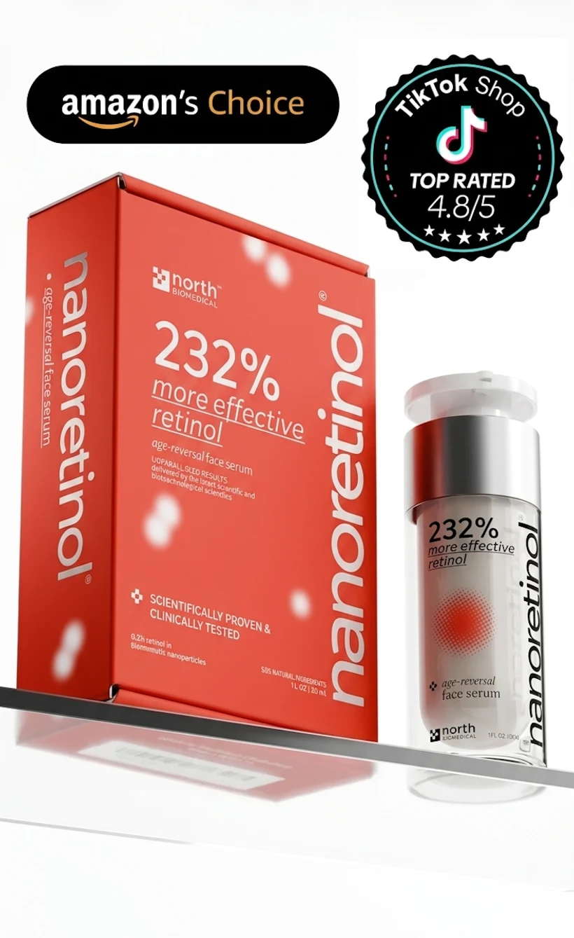
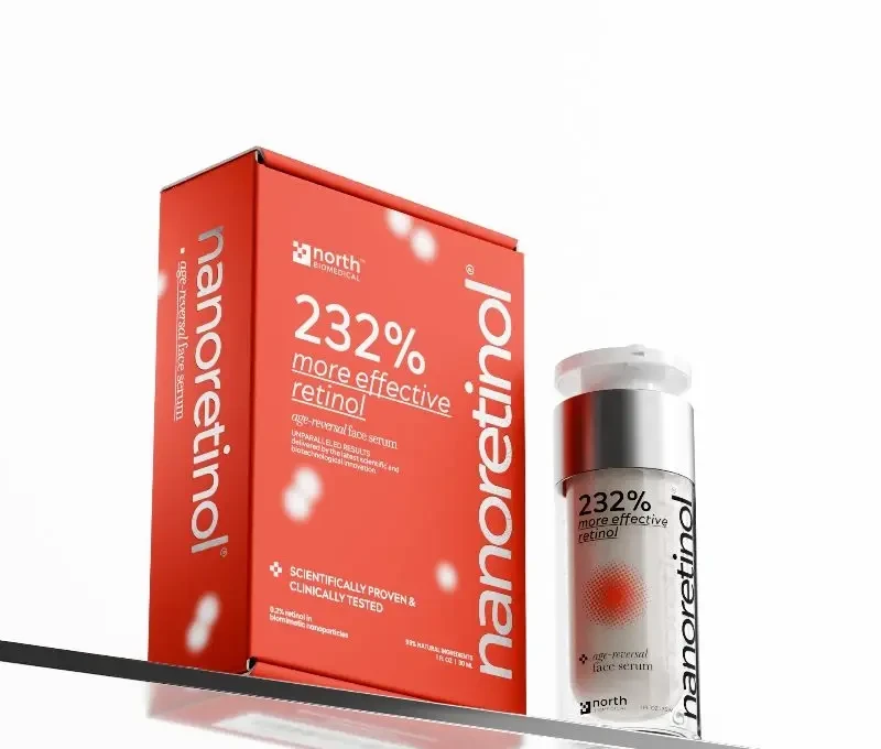
This is the central argument for advanced delivery technology. Nanoretinol encapsulates retinol in biomimetic lipid nanoparticles that are recognized by the skin as “self,” allowing penetration to the live cell layers where collagen synthesis occurs — without the barrier disruption that conventional formulations require. Clinical results: +232% more effective in collagen recovery, +61% increase in skin firmness at 56 days, with significantly reduced side effects versus conventional retinol [North Biomedical LLC. “Nanoretinol vs. Conventional Retinol: Efficacy in Collagen and Elastin Recovery.” Clinical Study Summary, 2024].
For something as structurally driven as nasolabial folds, maximizing how much retinol actually reaches the dermis matters.
Collagen-Stimulating Peptides: A Complementary Approach
Peptides have increasingly strong clinical evidence as a complement to retinoids. Signal peptides like palmitoyl pentapeptide-4 (Matrixyl) stimulate collagen types I, III, and IV synthesis independently of the retinoid pathway. GHK-Cu (copper peptides) influence gene expression across collagen, elastin, and glycosaminoglycan synthesis simultaneously.
Used alongside retinol, peptides address the same underlying deficit — insufficient collagen and elastin density — through a different mechanism. They’re generally well tolerated and have no meaningful conflict with retinoid use.
SPF: Stopping the Structural Damage From Compounding
UV radiation drives two simultaneous processes that worsen nasolabial folds: MMP-induced collagen degradation and solar elastosis — the abnormal accumulation and disorganization of elastin fibers under chronic UV exposure [3]. Without daily broad-spectrum sunscreen, any collagen-stimulating ingredient is fighting against ongoing UV-driven destruction.
Clinically, tretinoin at 0.025% produces results comparable to higher concentrations but with better tolerability.
The sunscreen for aging skin article covers the clinical evidence here in detail. The short version: no other single product in a skincare routine has stronger evidence for slowing the visible signs of facial aging than daily SPF.
A Note on Expression Lines vs. Volume Loss
Nasolabial folds have two distinct contributors that respond to different interventions:
- Expression-driven depth (from muscle contraction imprinting on skin) is addressed by improving skin quality — retinol, peptides, collagen density. This is the topical domain.
- Volume-driven depth (from fat redistribution and ligament laxity) responds to dermal fillers, fat transfer, or thread-based interventions. This is the procedural domain.
Many people have both contributors active. A realistic anti-aging routine addresses what topicals can do while being honest about when a consultation with a dermatologist or cosmetic physician is warranted.
The sunken cheeks and turkey neck articles address the structural aging changes that occur alongside nasolabial fold deepening.
What the Evidence Actually Supports for Daily Use
The combination supported by clinical research for improving skin quality across nasolabial folds:
Evening: Retinol (start slowly, build to nightly over four to six weeks). The goal is consistent collagen stimulation over months, not dramatic transformation in weeks.
Daytime: SPF 30+ broad-spectrum, applied daily regardless of cloud cover. Consider adding a niacinamide serum — it strengthens the skin barrier and may support hydration in the nasolabial area, where deeper furrows can create localized moisture loss.
Realistic timeline: Meaningful improvement in skin texture and fine line depth in eight to twelve weeks. Collagen density changes take longer — six months of consistent use to see structural benefit.
The folds will not disappear with a topical routine. But the speed at which they deepen, and the appearance of the skin across them, is genuinely influenced by the quality of collagen and elastin in the underlying dermis — and that is a target where retinoids, consistently applied, have decades of clinical evidence behind them.
References
-
Hong GW, Song S, Park SY, Lee SB, Wan J, Hu KS, Yi KH. “Why Do Nasolabial Folds Appear? Exploring the Anatomical Perspectives and the Role of Thread-Based Interventions.” Diagnostics (Basel). 2024;14(7):716. doi:10.3390/diagnostics14070716
-
Quan Y, Zhang T, Liu L, Han B, Cao Z, Shen Y. “Quantitative Assessment of Nasolabial Fold Characteristics Across Age Groups.” Aesthetic Surgery Journal Open Forum. 2025;7:ojaf075. doi:10.1093/asjof/ojaf075
-
Uitto J. “The role of elastin and collagen in cutaneous aging: intrinsic aging versus photoexposure.” Journal of Drugs in Dermatology. 2008;7(2 Suppl):s12–16. PMID:18404866
-
Kondo S, Ozawa N, Sakurai T. “The effect of degeneration of elastic fibres on loss of elasticity and wrinkle formation.” International Journal of Cosmetic Science. 2025;47(1):205–212. doi:10.1111/ics.13021
-
Ryu HS, Joo YH, Kim SO, Park KC, Youn SW. “Influence of age and regional differences on skin elasticity as measured by the Cutometer.” Skin Research and Technology. 2008;14(3):354–358. doi:10.1111/j.1600-0846.2008.00302.x
-
Mukherjee S, Date A, Patravale V, Korting HC, Roeder A, Weindl G. “Retinoids in the treatment of skin aging: an overview of clinical efficacy and safety.” Clinical Interventions in Aging. 2006;1(4):327–348. doi:10.2147/ciia.2006.1.4.327