
Solar Elastosis: What It Is, What Causes That Leathery Skin Texture, and What Reverses It
The science behind UV-induced elastin damage — and why tretinoin and retinol are the most evidence-based topical treatments
There’s a specific quality to certain kinds of aging skin that goes beyond wrinkles: a thick, slightly yellowish, leathery texture — the kind that doesn’t respond to moisturizer, doesn’t “firm up” with massage, and deepens over time regardless of what you put on top of it.
That texture has a name: solar elastosis. And it’s not simply accelerated aging. It’s a distinct pathological process — one that dermatologists can identify on biopsy, that differs mechanistically from the changes in chronologically aged skin, and that research suggests can, with the right intervention, partially reverse.
What Solar Elastosis Actually Is
In healthy skin, elastin is the protein that gives tissue its recoil — the property that allows you to pinch the back of your hand and watch it snap back. Elastin fibers are organized structures: they form on a scaffold of microfibrillar proteins including fibrillin-1, cross-linked by enzymes into functional elastic networks that run through the dermis in an orderly arrangement.
Solar elastosis disrupts this entirely. In photoaged skin, UV radiation drives the overproduction of tropoelastin — the precursor molecule that assembles into elastin — while simultaneously destroying the fibrillin-1 scaffolding needed to organize it [1]. The result is an accumulation of disorganized, insoluble elastin material that cannot function as elastic fiber. It piles up in the upper dermis as basophilic, amorphous clumps visible under microscopy — what pathologists call “elastotic material” [2]. That material accumulates one exposure at a time, which is the longer-term reason healing a sunburn properly is worth the patience it takes.
A second reinforcing mechanism makes the problem self-sustaining: UV-stimulated fibroblasts produce a protein called elafin, which acts as a protease inhibitor and effectively crystallizes the damaged elastin aggregates, preventing the skin’s natural degradation machinery from clearing them [3]. The elastotic material becomes locked in place.
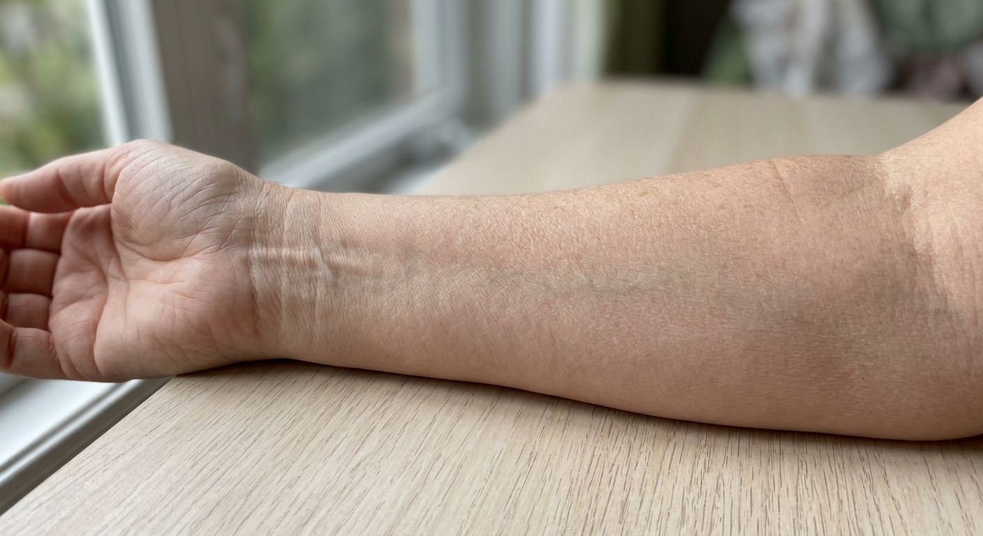
Clinically, solar elastosis produces the characteristic photoaged appearance: yellowish discoloration, coarse deep furrows, leathery texture, and the loss of the fine surface relief that makes younger skin look smooth.
How This Differs from Chronological Aging
The distinction between photoaging and chronological aging is not merely academic — it determines what will and won’t work as treatment.
In skin aging from time alone, elastin gradually depletes. Studies measuring elastin staining intensity across age decades in sun-protected skin show a slow, steady decline — from roughly 49% staining in early life to around 30% in the ninth decade [4]. Collagen diminishes similarly.
In sun-exposed skin, the pattern inverts. Elastin staining in photoaged facial skin actually increases with age — accumulating from around 57% to 75% over a lifetime [4]. The increase represents not functional elastin but the pathological elastotic material described above. Meanwhile, collagen fragmentation accelerates far earlier and more severely than in protected skin.
This is the paradox of solar elastosis: the tissue looks dense and thick because it is accumulating material, yet it is simultaneously less functional than younger, thinner skin. Normal aging produces atrophy; photoaging produces active, dysregulated accumulation of dysfunctional matrix alongside accelerated collagen destruction.
Elastin staining in photoaged facial skin actually increases with age — accumulating from around 57% to 75% over a lifetime.
An estimated 80% of visible facial aging is attributable to UV exposure rather than intrinsic aging — making photoaged skin a problem of cumulative UV dose far more than a function of chronological age [5]. That is why outdoor endurance athletes accumulate elastotic change so early, a pattern examined in our article on runner’s face.
Who Is Most Affected
Solar elastosis is dose-dependent: the more cumulative UV exposure, the more severe the elastosis. Research consistently shows elastosis severity tracks closely with lifetime UV dose rather than chronological age alone — individuals with equivalent biological ages but different sun exposure histories develop dramatically different degrees of photoaging.
Age itself is independently significant, since aging skin has diminished capacity to clear or repair elastotic material. Women over 40 are the primary clinical population for two compounding reasons: postmenopausal estrogen decline substantially reduces dermal collagen content — by roughly 30% in the first five years after menopause — leaving the dermis less capable of structural repair; and the sun-exposed areas where elastosis presents most prominently (perioral, neck, décolletage) are areas women are more likely to monitor and seek treatment for.
Women who grew up before broad sunscreen adoption and who have fair or light skin — both predictors of reduced UV protection — carry the highest lifetime elastosis burden from decades of cumulative exposure. Much of that dose was quieter than a beach day, because ordinary window glass passes a share of UVA while removing the UVB that would have warned them: see sunscreen indoors.
What the Evidence Says About Treatment
Topical Retinoids: The Most Supported Option
Tretinoin has the strongest evidence base of any topical intervention for solar elastosis, backed by over three decades of randomized controlled trial data.
The foundational mechanism was established in landmark research that demonstrated a single UV exposure upregulates collagenase (MMP-1), 92-kDa gelatinase (MMP-9), and stromelysin (MMP-3) in both the epidermis and dermis, increasing collagen fragmentation measurably in irradiated vs. non-irradiated skin. Critically, pre-treatment with tretinoin suppressed MMP induction by 70–80% — establishing retinoids’ pharmacological role not just in repairing existing damage but in blocking the enzyme cascade that causes it [5].
Further refinement of the mechanism identified MMP-12 — macrophage metalloelastase — as the primary enzyme responsible for degrading functional elastic fiber, with its expression strongly correlated with elastotic material accumulation in the upper dermis [6]. Topical retinoids suppress MMP-12 expression, partially restoring the balance between elastin production and clearance.
In terms of structural reversal: long-term studies using four or more years of tretinoin treatment document a 34% reduction in the dermal area occupied by elastosis. Shorter trials — 26 weeks or less — do not show elastosis reversal, consistent with the slow turnover rate of the accumulated elastotic material. Clinical results (smoother texture, reduced furrow depth) appear earlier than histological reversal, because retinoids also stimulate new collagen synthesis and epidermal thickening above the elastotic layer [5].
Vitamin C and Niacinamide as Adjuncts
These are not replacements for retinoids — they work upstream of the same UV-initiated pathways.
Vitamin C (ascorbic acid) is a potent antioxidant against UV-generated reactive oxygen species (ROS). Since ROS are the trigger for the MAPK and NF-kB signaling that drives MMP upregulation, topical vitamin C attenuates the cascade at its source. Clinical trials using topical vitamin C demonstrate histologically confirmed improvements in photoaged skin — improved texture and reduced wrinkles confirmed on biopsy — alongside objective skin brightening. It is most effective as a daily morning antioxidant, before UV exposure triggers the pathway.
For how each label number translates into UV actually reaching the dermis, compare SPF 30 vs 50.
Niacinamide (vitamin B3) operates through a distinct mechanism: it preserves cellular ATP stores under UV stress and enhances DNA repair capacity, attenuating the downstream ROS-mediated signaling that initiates MMP production and elastin disorganization [7].
The practical combination: vitamin C and niacinamide in the morning for protection; retinoid at night for repair.
Sunscreen: The Only True Prevention
No topical active can undo a lifetime of UV exposure rapidly. Sunscreen prevents the UV-initiated MMP cascade from occurring in the first place — making it the most efficient intervention relative to its cost and effort. Daily broad-spectrum SPF ≥30 is the foundational step for anyone with existing solar elastosis, both to prevent progression and to protect against further damage. Explore our guide to sunscreen for aging skin for evidence-based product selection. For how each label number translates into UV actually reaching the dermis, compare SPF 30 vs 50.
Where Nanoretinol Fits
The limiting factor for retinoid treatment is almost always tolerability, not efficacy. Tretinoin causes significant erythema, peeling, and barrier disruption at therapeutic concentrations — side effects severe enough that many patients discontinue before reaching the treatment duration (years, not weeks) needed for elastosis reversal.
Nanoretinol addresses this at the delivery level. Biomimetic lipid nanoparticles transport retinol through the epithelial barrier without disrupting it — the nanoparticles are structurally identical to skin cell membranes and absorbed passively, bypassing the barrier-disruption mechanism that drives tretinoin’s side effects. Clinical data show Nanoretinol achieves +232% greater collagen recovery efficiency than conventional retinol, with +61% improvement in skin firmness and +56% improvement in elasticity at 56 days, and with significantly reduced cytotoxicity.
For photoaged skin requiring years of consistent retinoid exposure, a formulation that can be used nightly without interruption due to irritation represents a meaningful clinical advantage. The delivery system makes the difference between a treatment that’s theoretically appropriate and one that’s practically sustainable.
Pair it with vitamin C serum in the morning and daily sunscreen to address the UV pathway from multiple angles — prevention upstream, repair downstream.
What to Expect
Solar elastosis that has accumulated over decades does not reverse in weeks. The realistic trajectory with consistent retinoid use is:
- Weeks 4–12: Improved texture at the surface; new collagen deposition begins; some brightening as melanin patterns normalize
- Months 3–6: Reduced furrow depth; smoother appearance; improved elasticity measurable clinically
- Year 1+: Gradual histological reduction in elastotic material; meaningful reversal requires sustained commitment
The skin you’re treating took decades to develop its current state. The reversal timeline reflects that — but the evidence is clear that meaningful change is achievable with the right approach and sufficient time.
Final Perspective
Solar elastosis is not an aesthetic problem you failed to prevent. It is a predictable biological outcome of cumulative UV exposure in an era before effective sun protection was understood or accessible. What dermatology now offers is a mechanistically coherent treatment path: suppress ongoing MMP activity, support new collagen synthesis, and over time, allow the skin’s natural clearance mechanisms to reduce the elastotic burden.
Retinoids sit at the center of that path because they address the mechanism — not just the appearance. How retinol builds collagen and why it remains the most evidence-backed topical for photoaged skin is worth understanding in depth before building your repair routine.
References
-
Widgerow AD, Napekoski K. “New approaches to skin photodamage histology — Differentiating ‘good’ versus ‘bad’ Elastin.” Journal of Cosmetic Dermatology. 2021;20(4):1098–1105. doi:10.1111/jocd.13865
-
Makino T, Kagoyama K, Murabe C, Nakamura T, Shimizu T. “Association of development of solar elastosis with increased expression of fibrillin-1, LTBP-2 and fibulin-4 in combination with decreased expression of LTBP-4.” Acta Dermato-Venereologica. 2021;101:adv00388. doi:10.2340/00015555-3738
-
Pain S, Berthélémy N, Naudin C, Degrave V, André-Frei V. “Understanding solar skin elastosis — cause and treatment.” Journal of Cosmetic Science. 2018;69(3):175–185. PMID: 30052192
-
El-Domyati M, Attia S, Saleh F, et al. “Intrinsic aging vs. photoaging: a comparative histopathological, immunohistochemical, and ultrastructural study of skin.” Experimental Dermatology. 2002;11(5):398–405. doi:10.1034/j.1600-0625.2002.110501.x
-
Fisher GJ, Wang ZQ, Datta SC, et al. “Pathophysiology of premature skin aging induced by ultraviolet light.” New England Journal of Medicine. 1997;337(20):1419–1428. doi:10.1056/NEJM199711133372003
-
Pittayapruek P, Meephansan J, Prapapan O, Komine M, Ohtsuki M. “Role of matrix metalloproteinases in photoaging and photocarcinogenesis.” International Journal of Molecular Sciences. 2016;17(6):868. doi:10.3390/ijms17060868
-
Quan T, Little E, Quan H, Voorhees JJ, Fisher GJ. “Elevated matrix metalloproteinases and collagen fragmentation in photodamaged human skin: impact of altered extracellular matrix microenvironment on dermal fibroblast function.” Journal of Investigative Dermatology. 2013;133(5):1362–1366. doi:10.1038/jid.2012.509