
Crow's Feet: What Causes Them and How to Actually Treat Them
The science behind lateral canthal lines, why the eye area ages first, and evidence-based treatments that deliver real results
The lines that fan outward from the corners of your eyes have one of the most evocative names in skincare. Crow’s feet — or lateral canthal lines, if you prefer the clinical term — are among the very first visible signs of skin aging. Most people notice them in their early thirties, though the structural changes that produce them begin much earlier.
Understanding why crow’s feet appear requires looking at what makes the eye area fundamentally different from the rest of the face. This is not just thin skin and squinting. It is a convergence of anatomical factors that make the periorbital region uniquely vulnerable to aging.
Why the Eye Area Ages First
The skin around the eyes is the thinnest on the entire body — roughly 0.5 mm compared to 2 mm on the cheeks [3]. This matters because thinner skin contains fewer layers of collagen and elastin fibers, the structural proteins that keep skin firm and resilient. With less structural reserve, the periorbital area shows damage earlier and more visibly.
Shuster et al. established that skin thickness, collagen content, and density all decline with age, with the rate of decline varying by body site [3]. The eye area, starting from the thinnest baseline, reaches a visible threshold of damage before virtually any other region.
Compounding this is the orbicularis oculi — the circular muscle that encircles the eye and controls blinking, squinting, and smiling. Humans blink approximately 15,000 to 20,000 times per day. Each blink contracts the orbicularis oculi, and each contraction creases the overlying skin. Over decades, these repetitive micro-folds gradually etch permanent lines into skin that has progressively less collagen to resist them.
Piérard-Franchimont et al. demonstrated that the tensile properties of skin collagen deteriorate measurably with age [6]. Collagen fibers become stiffer and less elastic, meaning the skin around the eyes loses its ability to snap back after each contraction. The crease that once vanished the moment you stopped smiling eventually stays.
The Collagen Connection
Collagen loss is the central driver of crow’s feet progression. After age 20, the skin loses roughly 1% of its collagen per year. By 50, you have lost approximately 30% of the collagen you had at your peak.
Varani et al. found that naturally aged human skin shows both decreased collagen production and elevated levels of matrix metalloproteinases (MMPs) — enzymes that actively break down existing collagen [4]. This creates a double deficit: less new collagen is being built while more existing collagen is being degraded.
UV exposure accelerates this process dramatically. Photoaged skin shows collagen damage far exceeding what chronological aging alone would produce [2]. Since the periorbital area receives significant sun exposure — people rarely apply adequate sunscreen around the eyes — crow’s feet often represent a combination of intrinsic aging and cumulative photodamage.
Elastin loss follows a parallel trajectory. The elastic fibers that allow skin to stretch and recoil degrade with age and UV exposure, contributing to the permanent creasing pattern that defines crow’s feet. If you want to understand the broader relationship between retinol and collagen production, the science is well-established and directly applicable to the eye area.
By 50, you have lost approximately 30% of the collagen you had at your peak.
Treatment Options That Actually Work
Topical Retinoids: The Gold Standard for At-Home Treatment
Retinoids remain the most evidence-backed topical treatment for crow’s feet. The mechanism is well understood: retinoids bind to nuclear receptors in skin cells, upregulating collagen synthesis while suppressing the MMPs that degrade it [1].
Mukherjee et al. conducted a comprehensive review of retinoids in skin aging and confirmed their efficacy in reducing fine lines, improving skin texture, and stimulating collagen production [1]. The evidence spans decades of clinical trials and histological studies.
Kang et al. showed that even retinol — the over-the-counter form of vitamin A — induces meaningful biological changes in human skin, including epidermal thickening and increased expression of cellular retinoid binding proteins [5]. Importantly, these changes occurred without the irritation typically associated with prescription-strength tretinoin.
Bhawan et al. conducted histologic studies of tretinoin-treated photodamaged skin and found increased collagen deposition in the upper dermis, reduced melanin irregularities, and normalized epidermal architecture [2]. These structural changes correspond directly to the visible reduction in fine lines and improved skin texture that users report.
The challenge with retinoids around the eyes is tolerability. Conventional retinol formulations can cause significant irritation in the thin, sensitive periorbital area — redness, peeling, and dryness that sometimes make the eye area look worse before it looks better. This is where formulation science becomes critical. For more detail on using retinol safely around the eyes, delivery method matters as much as the active ingredient.
Peptides and Growth Factors
Peptide serums represent a gentler complementary approach. Signal peptides like palmitoyl pentapeptide-4 (Matrixyl) stimulate fibroblasts to produce collagen, though the evidence base is smaller than for retinoids, and a peptide eye cream is the usual way to apply them around the eyes, while a collagen eye cream mostly adds hydration. They work best as part of a comprehensive routine rather than as standalone treatments.
Growth factor serums derived from stem cell conditioned media show promise in early studies for improving periorbital skin quality, though long-term data is still limited.
Professional Treatments
For moderate to severe crow’s feet, professional interventions include botulinum toxin injections (which relax the orbicularis oculi muscle to reduce dynamic lines), laser resurfacing (which stimulates collagen remodeling through controlled thermal injury), and radiofrequency microneedling (which delivers heat to the dermal layer to trigger new collagen production).
These treatments are effective but come with costs, downtime, and the need for maintenance sessions. Many dermatologists recommend combining professional treatments with a strong at-home topical routine — retinoids as the foundation — for the best sustained results.
Clinical data shows Nanoretinol® delivers +232% more effective collagen recovery and +73% more effective elastin recovery compared to conventional retinol, while being significantly gentler.
Sunscreen: The Non-Negotiable
No treatment strategy for crow’s feet is complete without daily broad-spectrum SPF 30 or higher. UV radiation is the primary external driver of collagen degradation around the eyes [2]. Wearing sunglasses provides additional protection by reducing squinting and shielding the periorbital skin from direct UV exposure. Getting the filter itself to stay put on that skin is its own problem — see sunscreen around the eyes.
Why Formulation Matters for the Eye Area
The skin around the eyes poses a formulation challenge. It needs retinoid delivery that is effective enough to stimulate collagen production but gentle enough to avoid overwhelming the thinnest, most sensitive skin on the face. Many people abandon retinol eye creams because conventional formulations trigger too much irritation in this delicate zone.
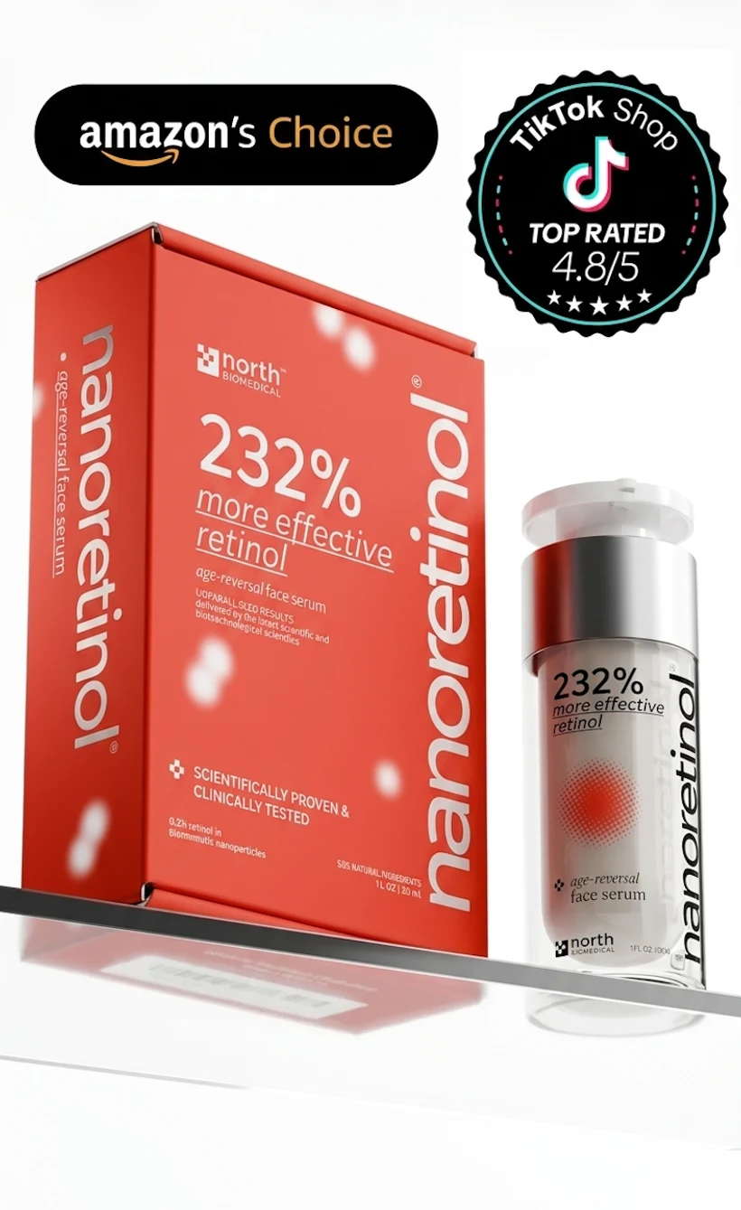
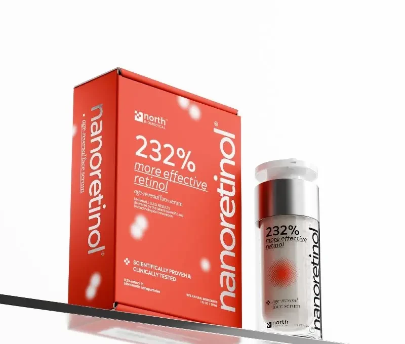
This is precisely the problem that lipid nanoparticle encapsulation was designed to solve. Nanoretinol® by North Biomedical® uses biomimetic lipid nanoparticles to deliver retinol at 0.2% concentration in a water-based gel. The encapsulation provides controlled, gradual release — meaning the retinol reaches target cells in the dermis without overwhelming the thin periorbital epidermis.
Clinical data shows Nanoretinol® delivers +232% more effective collagen recovery and +73% more effective elastin recovery compared to conventional retinol, while being significantly gentler [7]. For the eye area, where choosing the right eye cream can make or break your results, this combination of enhanced efficacy and reduced irritation is particularly relevant.
The 99% natural ingredient profile and water-based gel texture also mean no heavy oils sitting in the eye area — a common complaint with traditional retinol creams that can migrate into the eyes and cause stinging.
Building a Crow’s Feet Routine
An effective routine for treating crow’s feet does not need to be complicated:
- Morning: Gentle cleanser, antioxidant serum (vitamin C), moisturizer, SPF 30+ sunscreen, sunglasses
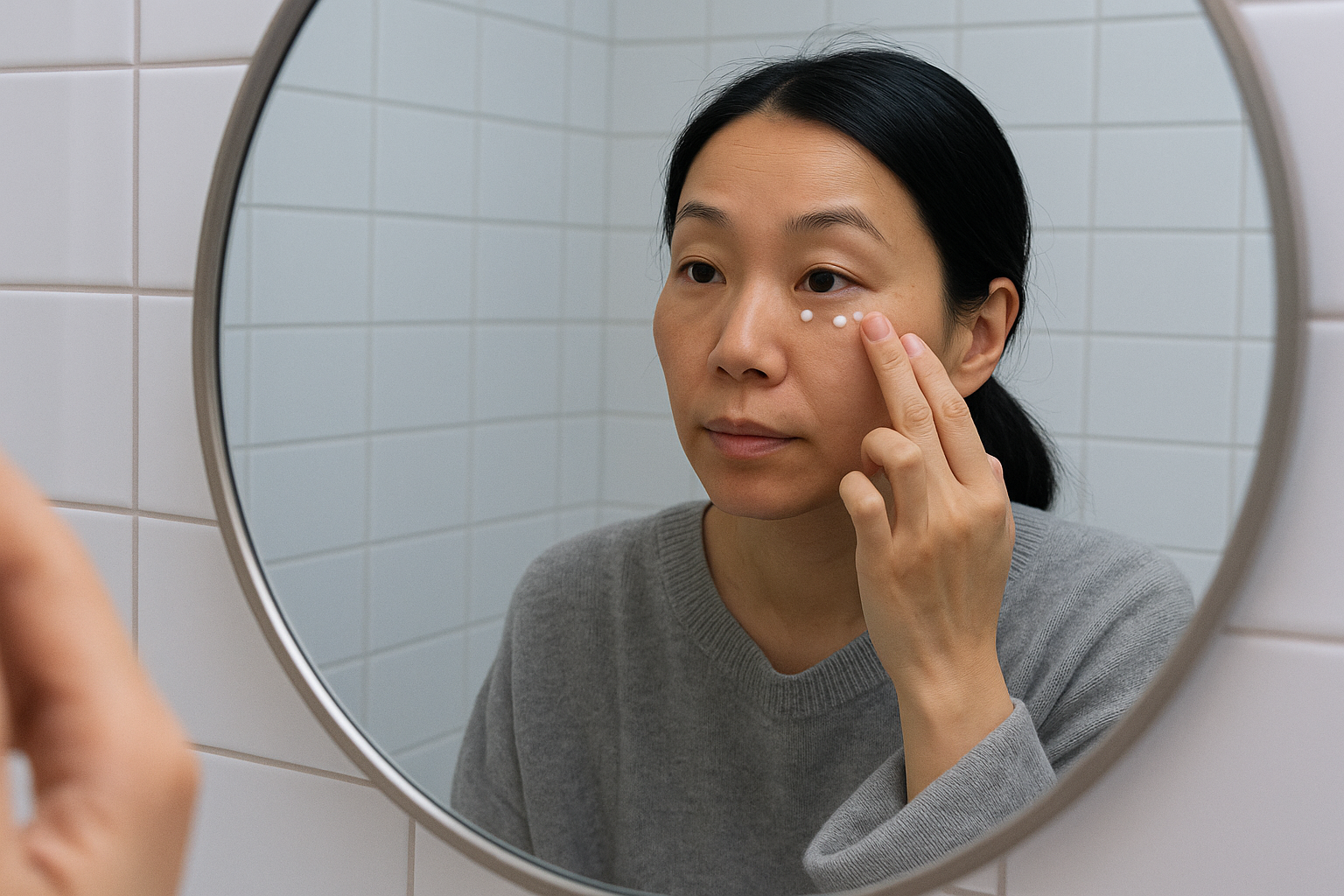
- Evening: Gentle cleanser, retinoid treatment (applied carefully to the orbital bone area, not the eyelid), hydrating eye cream or moisturizer
Start with retinoid application every other night and increase to nightly as tolerated. If you have concerns about retinol thinning the skin — a common myth — the evidence shows the opposite: retinoids actually thicken the epidermis over time [5].
Results from topical retinoids typically become visible after 8 to 12 weeks of consistent use, with continued improvement over 6 to 12 months. The biology of collagen synthesis requires patience. Lines that took decades to form will not vanish in days — but they can measurably improve with the right approach.
Crow’s feet are a universal sign of a life spent expressing emotion. Treating them effectively means understanding the unique biology of the eye area and choosing treatments that work with that biology rather than against it. The science is clear: consistent retinoid use, sun protection, and proper formulation are the evidence-based foundation for reducing lateral canthal lines. The approach for addressing forehead wrinkles follows similar principles, since the same collagen biology applies across the face.
References
-
Mukherjee S, Date A, Patravale V, Korting HC, Roeder A, Weindl G. Retinoids in the treatment of skin aging: an overview of clinical efficacy and safety. Clin Interv Aging. 2006;1(4):327-348. PMID: 18046911
-
Bhawan J, Gonzalez-Serva A, Nehal K, et al. Effects of tretinoin on photodamaged skin. A histologic study. Arch Dermatol. 1991;127(5):666-672. PMID: 2024984
-
Shuster S, Black MM, McVitie E. The influence of age and sex on skin thickness, skin collagen and density. Br J Dermatol. 1975;93(6):639-643. doi:10.1111/j.1365-2133.1975.tb05113.x
-
Varani J, Dame MK, Rittie L, et al. Vitamin A antagonizes decreased cell growth and elevated collagen-degrading matrix metalloproteinases and stimulates collagen accumulation in naturally aged human skin. J Invest Dermatol. 2000;114(3):480-486. doi:10.1046/j.1523-1747.2000.00876.x
-
Kang S, Duell EA, Fisher GJ, et al. Application of retinol to human skin in vivo induces epidermal hyperplasia and cellular retinoid binding proteins characteristic of retinoic acid but without measurable retinoic acid levels or irritation. J Invest Dermatol. 1995;105(4):549-556. doi:10.1111/1523-1747.ep12323445
-
Piérard-Franchimont C, Colige A, Piérard GE. Tensile properties and contraction of skin collagen in relationship to age. Mech Ageing Dev. 1998;100(1):1-7. doi:10.1016/S0047-6374(97)00114-2
-
North Biomedical LLC. “Nanoretinol® vs. Conventional Retinol: Efficacy in Collagen and Elastin Recovery.” Clinical Study Summary, 2024.