
Milia Under Eyes: Why Those Tiny White Bumps Form and How to Actually Get Rid of Them
The dermatology-backed guide to milia removal — from periocular anatomy to retinoid treatment
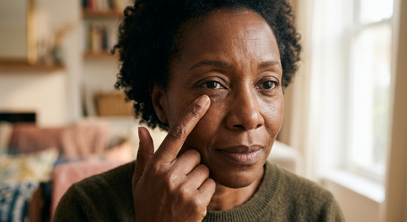
You notice them in certain light — tiny, pearl-white bumps nestled just below the lower lash line, firm to the touch, stubbornly fixed no matter how much you exfoliate. Squeezing does nothing. Scrubbing does nothing. They look like whiteheads but they don’t behave like them.
They’re milia. And they’re one of the more consistently misunderstood skin conditions in adult dermatology. The good news: once you understand what they actually are — and why your skin makes them — the path to clearing them becomes much clearer.
What Milia Actually Are
Milia are benign keratin-filled cysts: tiny enclosed capsules of the same structural protein that forms your nails and hair, trapped beneath a closed layer of skin. Histologically, each milium consists of a small cavity lined by stratified squamous epithelium, packed with compact keratinous material [1].
They are not whiteheads. A whitehead — a closed comedone — is a plugged hair follicle containing a mix of keratin and sebum. It has the potential to be expressed because it has a connection, however blocked, to the skin’s surface. A milium has no external opening. The keratin inside has nowhere to go, which is why these bumps can persist for months or years without intervention.
Milia fall into two categories with distinct origins. Primary milia form spontaneously, arising from the infundibular zone of vellus hair follicles. Immunohistochemical analysis using keratin markers traces their origin to the outermost layer of the hair bulge of the outer root sheath — the zone containing follicular stem cells [2]. Secondary milia form in response to skin disruption: burns, laser resurfacing, chronic topical corticosteroid use, or blistering diseases. In secondary milia, roughly 75% connect to eccrine sweat duct units rather than hair follicles [3].
Why the Eye Area Is a Prime Location
The skin around the eyes is the thinnest on the human body — approximately 0.5 mm, compared to 2 mm on the back. It is also densely populated with vellus hair follicles, the exact structures from which primary milia originate [1].
This anatomy creates ideal conditions for keratin cysts. Follicular channels sit close to the surface, but the overlying skin is too thin and too dry to facilitate normal desquamation. Unlike the nose or forehead — where sebum keeps follicular ducts patent — the periocular area is relatively sebum-poor. Keratin that would normally shed as part of regular epidermal turnover instead accumulates in a closed pocket and compacts over weeks or months into the firm, visible bump you see.
Who Gets Milia — and Why Women Over 40 Are Particularly Susceptible
Milia are not exclusively a midlife problem. Around 40–50% of healthy newborns develop congenital milia that resolve spontaneously within weeks. But the clinical pattern in adult women over 40 is consistent enough that dermatologists recognize a distinct clustering [1].
In secondary milia, roughly 75% connect to eccrine sweat duct units rather than hair follicles.
A specific subtype — milia en plaque — shows a strong female predominance and typically develops between the fourth and seventh decades of life. The periocular region is the most commonly reported location for this variant.
Several biological converge in midlife:
Slowing epidermal turnover. After 40, the natural cell-to-surface journey slows from roughly 28 days in young skin to 45–60 days or longer. Slower turnover means trapped keratin has more time to compact into a fixed cyst before reaching the surface.
Postmenopausal skin changes. Declining estrogen reduces epidermal thickness and barrier function, impairs desquamation, and compromises follicular duct patency — all of which increase the likelihood of keratin being retained rather than shed.
Cumulative photoaging. Years of UV exposure alter follicular architecture and keratinization patterns in the periocular zone specifically, disrupting the normal exit pathway for keratin.
None of these factors is dramatic on its own. Together, they shift the balance from “skin sheds keratin normally” to “skin traps keratin.”
What Actually Works for Milia Removal
Professional Extraction
The gold standard for existing milia is mechanical extraction by a trained clinician. A lancet or fine scalpel creates a micro-incision through the outer skin; the keratin contents are then expressed using a comedone extractor or sterile curette [3].
Do not attempt this at home. The skin around the eyes is not only the thinnest on the face — it’s also in close proximity to the globe. An imprecise incision can cause scarring. An unsanitary one introduces infection risk in tissue that heals less predictably than elsewhere on the face.
Around 40–50% of healthy newborns develop congenital milia that resolve spontaneously within weeks.
Topical Retinoids
For prevention and for those who develop milia recurrently, topical retinoids — retinol and prescription tretinoin — are the most evidence-based topical option [1, 3]. The mechanism is well-characterized: retinoids bind to retinoic acid receptors (RAR) in keratinocyte nuclei, activating gene programs that accelerate cell turnover, loosen corneocyte cohesion, and normalize follicular keratinization [4]. By speeding up the shedding of surface cells and keeping follicular channels clear, retinoids prevent the static accumulation that allows milia to form.
Prescription tretinoin, at 0.025% to 0.05%, is most commonly used for this purpose. Over-the-counter retinol requires conversion to retinoic acid through two enzymatic steps, making its effect slower but still meaningful for prevention [4].
Laser and Energy-Based Treatments
For milia en plaque or cases resistant to other treatments, erbium:YAG laser ablation has demonstrated success with minimal collateral damage — particularly relevant near the eyes, where the CO2 laser’s thermal spread poses a greater scarring risk. Electrodesiccation, cryotherapy, and photodynamic therapy are also used depending on lesion depth and skin type.
The Eye Area Challenge: Why Delivery Method Matters
The periocular zone is the hardest area to treat with conventional retinoids. Tretinoin’s standard mechanism — disrupting the lipid barrier to penetrate skin — causes erythema and peeling even on the broader face. Around the eyes, where skin is thinnest and most reactive, the same concentrations that work elsewhere frequently cause intolerable irritation. Many women stop before they achieve the prevention benefit.
This is where retinol delivery technology changes the calculus. Nanoretinol uses biomimetic lipid nanoparticles — particles structurally identical to skin cell membranes — to transport retinol through the epithelial barrier without disrupting it [4]. Instead of forcing entry, the nanoparticles are recognized as “self” by skin cells and absorbed passively, delivering retinol to target tissue without triggering the barrier-disruption cascade that causes irritation.
In clinical testing, Nanoretinol achieved a +61% increase in skin firmness and +56% increase in elasticity in 56 days, supported by +232% greater collagen recovery efficiency than conventional retinol — with significantly reduced cytotoxicity and minimal side effects. For the delicate periocular area, tolerability isn’t a secondary concern. It’s the primary one. Learn how retinol around the eyes differs from general facial use — and what to watch for in any periocular retinoid routine.
Managing Expectations
Established milia — particularly those that have mineralized over months — require physical extraction or sustained retinoid use over weeks to months for visible clearing. They do not dissolve quickly regardless of the approach.
For prevention: consistent retinoid use combined with gentle chemical exfoliation (lactic or mandelic acid, both better tolerated around the eyes than glycolic acid) reduces recurrence significantly. Avoid heavy occlusive eye creams in the immediate periocular zone — they can further impede follicular clearance. Daily SPF protects the follicular architecture that UV damage progressively impairs. If you’re managing eyelid wrinkles and dark circles under eyes simultaneously, a well-formulated retinoid addresses multiple concerns from the same area with consistent, long-term use.
If milia multiply rapidly, appear across a large area suddenly, or coincide with a new medication, consult a dermatologist — secondary milia can occasionally signal an underlying condition worth investigating.
References
-
Berk DR, Bayliss SJ. “Milia: a review and classification.” Journal of the American Academy of Dermatology. 2008;59(6):1050–1063. doi:10.1016/j.jaad.2008.07.034
-
Kurokawa I, Kakuno A, Tsubura A. “Milia may originate from the outermost layers of the hair bulge of the outer root sheath: A case report.” Oncology Letters. 2016;12(6):5190–5192. doi:10.3892/ol.2016.5335
-
Gallardo Avila PP, Mendez MD. “Milia.” StatPearls [Internet]. Treasure Island, FL: StatPearls Publishing; 2024 Jan. PMID: 32809316
-
Zasada M, Budzisz E. “Retinoids: active molecules influencing skin structure formation in cosmetic and dermatological treatments.” Advances in Dermatology and Allergology. 2019;36(4):392–397. doi:10.5114/ada.2019.87443