
Sebaceous Hyperplasia: What Those Bumpy Yellow Spots on Your Face Actually Are
A dermatology-backed guide to sebaceous hyperplasia treatment, causes, and why it's so often misdiagnosed
You notice a cluster of small, yellowish, slightly shiny bumps on your forehead or cheeks — not quite whiteheads, not quite pimples, with a faint dimple at the center you can’t quite explain. Your instinct might be to treat them like acne. But they don’t respond the same way. That’s because they aren’t acne.
What you’re likely looking at is sebaceous hyperplasia — an enlargement of the oil-producing glands embedded in your skin. It’s one of the more common dermatological findings in middle-aged adults, frequently biopsied to rule out something more serious, and consistently under-explained to the patients who have it.
What Sebaceous Hyperplasia Is
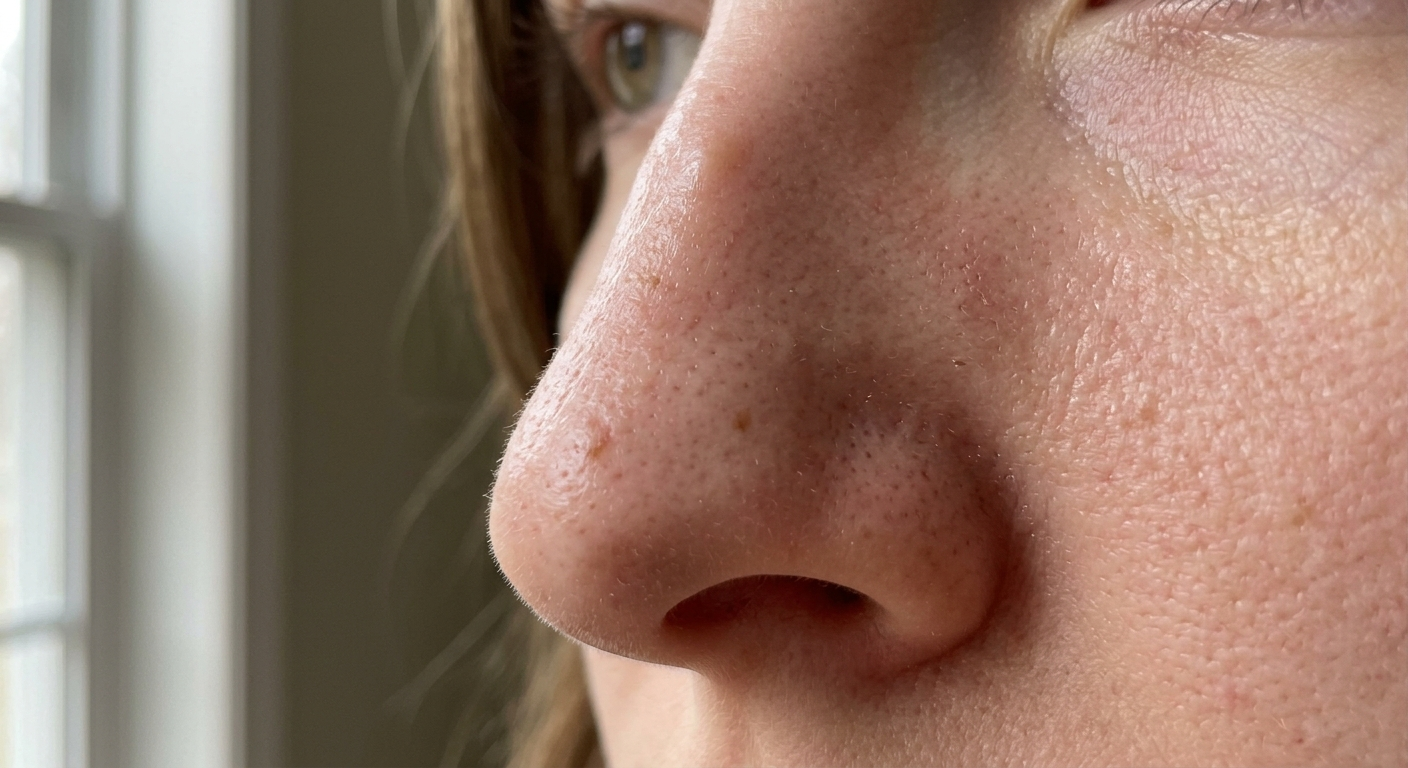
Sebaceous glands are the microscopic oil-producing structures that open into hair follicles across most of the face and body. In sebaceous hyperplasia (SGH), these glands don’t malfunction — they enlarge. Each lesion consists of a single expanded sebaceous gland with multiple mature lobules packed around a widened central duct [1]. Clinically, this produces the characteristic appearance: a 2–9 mm yellowish papule, soft to the touch, with a central umbilication — that small central dimple — through which sebum can sometimes be gently expressed.
That central depression is the key diagnostic feature. Whiteheads lack it. Milia lack it. The condition most frequently confused with SGH — basal cell carcinoma — also lacks it [1].
Sebaceous hyperplasia affects roughly 1% of the general adult population — but that number climbs to 10–16% among organ transplant patients taking cyclosporine A, a drug known to alter sebocyte biology [1]. This association reveals something important: SGH is not primarily a sebum-output problem. It’s a cellular turnover problem.
The Aging Paradox of Oil Glands
Here’s where sebaceous hyperplasia becomes counterintuitive. You might expect enlarged oil glands to be associated with high sebum production — oily skin, hormonal surges, youth. In reality, SGH is most common in the fifth and sixth decades of life, precisely when sebum production is declining [1, 4].
The mechanism works like this: sebocytes (the cells within sebaceous glands) are continuously born, mature, and shed — a turnover process driven partly by androgen signaling. As people age, that turnover rate slows. Sebocytes cycle through the gland more slowly, accumulating rather than being shed. The gland expands structurally without increasing its functional output [1].
Sebaceous glands are among the most androgen-sensitive structures in the body. They express the full complement of enzymes — 5-alpha reductase type I, 3-beta-hydroxysteroid dehydrogenase, 17-beta-hydroxysteroid dehydrogenase — needed to locally convert circulating androgens into dihydrotestosterone (DHT), the most potent driver of sebocyte activity [4]. But direct androgen excess doesn’t seem to be the cause of SGH. A controlled study comparing circulating androgen levels in women with SGH against matched controls found no statistically significant difference [1]. The problem appears to lie in glandular dynamics — a slowdown in how efficiently cells move through the sebaceous gland — rather than in excess stimulation from androgens circulating systemically.
What you’re likely looking at is sebaceous hyperplasia — an enlargement of the oil-producing glands embedded in your skin.
In women, this transition correlates with perimenopause, when falling estrogen shifts the hormonal environment and alters skin cell kinetics across multiple pathways simultaneously.
Why It Gets Misdiagnosed
Sebaceous hyperplasia is biopsied specifically to exclude basal cell carcinoma (BCC) far more often than most skin conditions. The clinical overlap is real: both present as yellowish, pearlescent papules on sun-exposed facial skin of older adults, sometimes with visible surface vessels.
The distinguishing features of SGH:
- Central dell: A visible umbilication at the center, often with a small pore
- Sebum expression: Gentle lateral pressure sometimes expresses a small amount of oily material
- Grouped lobular pattern: Under dermoscopy, the classic “morulae” pattern — aggregated white-yellow lobules surrounding a central ostium — with radially arranged “crown vessels” that stop at the lesion edge and do not cross the center [1]
BCC shows none of these features. Its pearlescent quality comes from basaloid proliferation, and dermoscopy reveals arborizing vessels, leaf-like structures, and blue-gray dots — patterns specific to carcinoma.
The other common misdiagnosis is milia, which share the white-to-yellow color but are harder, smaller, and lack both the central dimple and the lobular grouping visible on close examination.
What Actually Works for Treatment
Sebaceous hyperplasia is benign. Treatment is cosmetic, not medically necessary — but the cosmetic impact is real, particularly as lesions accumulate in number and size over years.
Oral isotretinoin is the most extensively studied systemic treatment. A clinical trial of 20 patients at 1 mg/kg/day for two months reduced mean lesion count from 24 to 2, with only 4 lesions observed at two-year follow-up [2]. The catch: sebaceous hyperplasia frequently recurs after isotretinoin is discontinued, and the dose required for SGH is similar to that for severe acne — with corresponding side effects.
Photodynamic therapy (PDT) combining topical aminolevulinic acid with pulsed dye laser (595 nm) has demonstrated clinical efficacy. In a prospective trial, this combination approach outperformed laser-alone controls, with side effects limited to transient erythema and focal crusting [3]. PDT selectively targets the sebaceous gland — aminolevulinic acid preferentially accumulates in sebocyte-rich tissue — making it an elegant mechanism for this specific problem.
Topical tretinoin (starting at 0.025% and escalating) is most commonly used clinically.
Laser and light therapies reviewed systematically include the pulsed dye laser, CO2 laser, argon laser, and 1720 nm wavelength devices that specifically target sebaceous lipids. Combination approaches achieve higher clearance rates, though individual results vary significantly by lesion count and skin phototype.
Electrodesiccation and cryotherapy remain reliable conventional options for isolated lesions, with good cosmetic outcomes when performed precisely.
Topical Retinoids: Prevention and Maintenance
Topical retinoids occupy a specific role in managing sebaceous hyperplasia — not as acute clearance agents, but as long-term preventive tools.
The mechanism is well-established from the isotretinoin literature and translates to topical application: retinoids act on retinoic acid receptors (RAR) within sebocytes to inhibit their proliferation, suppress lipid synthesis, and normalize the cellular turnover that drives gland enlargement [4]. Topical tretinoin (starting at 0.025% and escalating) is most commonly used clinically. Over-the-counter retinol exerts a similar but milder effect through the same pathway, requiring enzymatic conversion to retinoic acid before it becomes biologically active.
The practical effect of consistent topical retinoid use: slower accumulation of new lesions, reduced gland size over months of use, and secondary benefits for photoaging that often concern this same demographic. For maintenance, the tolerability of the retinoid formula matters enormously — sebaceous hyperplasia tends to appear on facial skin that may already be reactive or in areas where the complexion is sensitive.
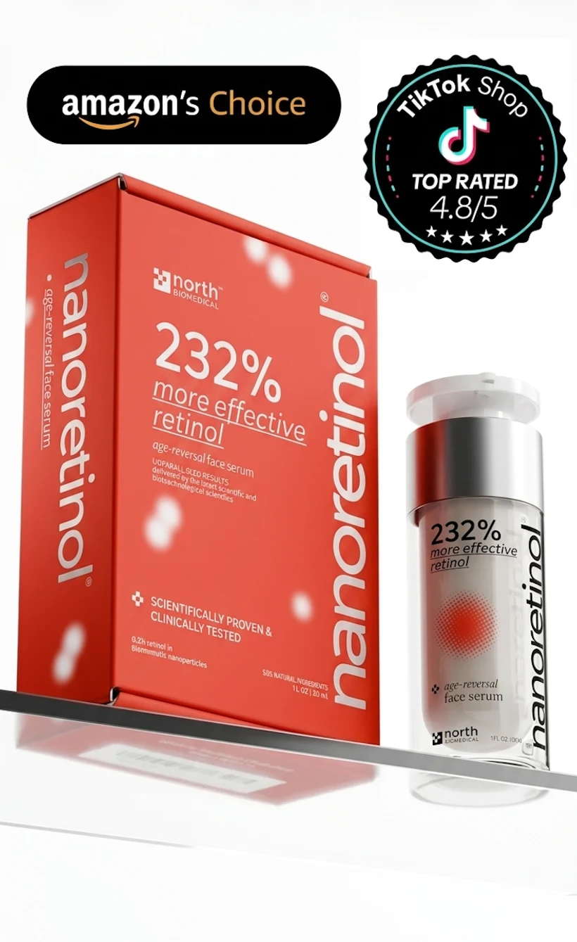
Nanoretinol’s biomimetic nanoparticle delivery addresses this directly. Rather than penetrating skin by disrupting its lipid barrier — the mechanism that causes redness and peeling in conventional retinol formulations — Nanoretinol’s particles are recognized as structurally identical to skin cell membranes and absorbed passively. The result is effective retinol delivery to the sebaceous gland level without barrier disruption-related irritation. Clinical testing showed +232% greater collagen recovery efficiency versus conventional retinol, with significantly reduced cytotoxicity. For a condition that requires months-to-years of maintenance retinoid use, tolerability without compromising efficacy is the core advantage.
If you’re also managing enlarged pores or oily skin texture alongside sebaceous hyperplasia, a retinoid-centered routine addresses all three concerns through the same mechanism — normalization of sebocyte biology and epidermal turnover. For foundational knowledge on how niacinamide benefits complement retinoid use by regulating sebum production, that pairing is particularly relevant for SGH management.
When to See a Dermatologist
Any lesion on the face of an adult over 40 that is pearlescent, yellowish, or vascular and does not have the characteristic central dell of SGH deserves clinical evaluation. Basal cell carcinoma is common on sun-exposed facial skin in this demographic and can mimic sebaceous hyperplasia visually.
Similarly, if lesions multiply rapidly, appear in unusual locations, or emerge after starting a new medication, consult a dermatologist. Cyclosporine A, certain HIV antiretrovirals, and chronic corticosteroid use are all documented triggers for secondary sebaceous hyperplasia — and identifying the cause opens different management options.
For typical SGH — gradual accumulation on the nose, cheeks, and forehead in middle age — the combination of in-office treatment for existing lesions and consistent topical retinoid use for prevention is the most evidence-aligned long-term approach.
References
-
Farci F, Rapini RP. “Sebaceous Hyperplasia.” StatPearls [Internet]. Treasure Island, FL: StatPearls Publishing; 2023 Sep. https://www.ncbi.nlm.nih.gov/books/NBK562148/
-
Tagliolatto S, Santos Neto OO, Alchorne MMA, Enokihara MY. “Sebaceous hyperplasia: systemic treatment with isotretinoin.” Anais Brasileiros de Dermatologia. 2015;90(2):211–215. doi:10.1590/abd1806-4841.20153192
-
Alster TS, Tanzi EL. “Photodynamic therapy with topical aminolevulinic acid and pulsed dye laser irradiation for sebaceous hyperplasia.” Journal of Drugs in Dermatology. 2003;2(5):501–504. PMID: 14558397
-
Thody AJ, Shuster S. “Control and function of sebaceous glands.” Physiological Reviews. 1989;69(2):383–416. doi:10.1152/physrev.1989.69.2.383