Age Spots on Hands: What They Really Are and How to Fade Them
The dermatology behind solar lentigines, why hands show sun damage first, and proven treatments to reduce and prevent dark spots
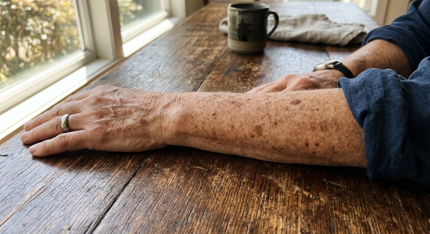
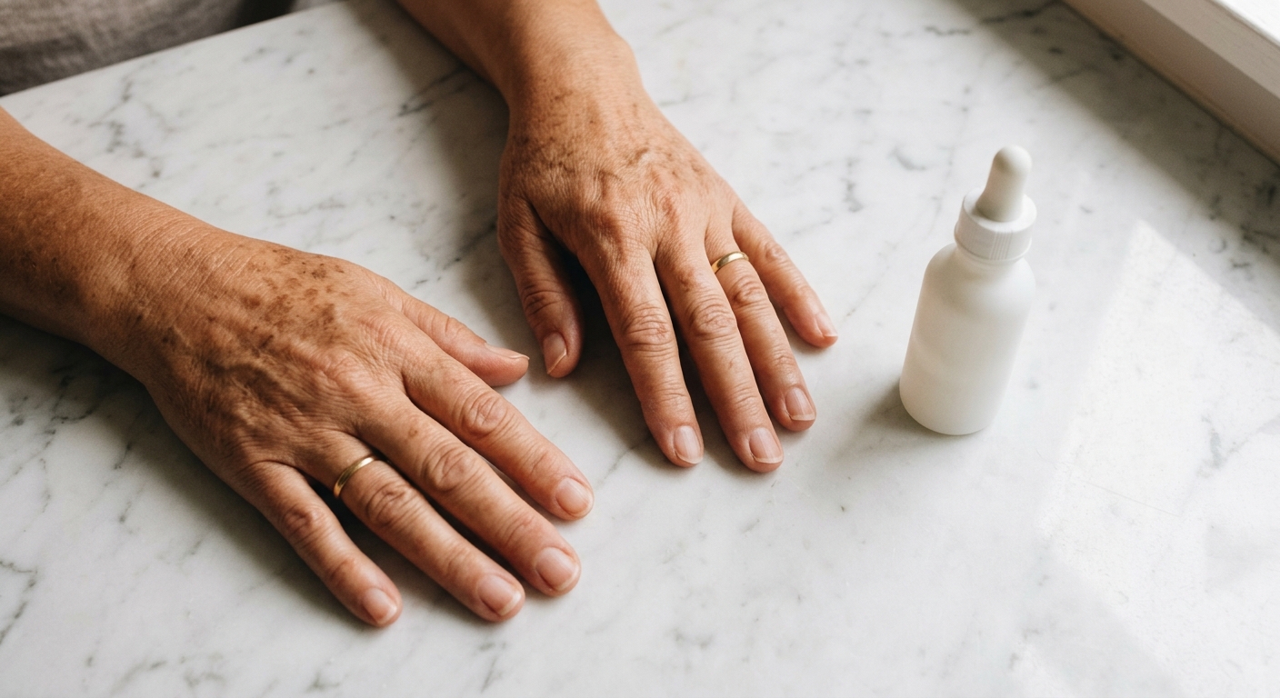
The flat brown spots that appear on the backs of hands after decades of sun exposure go by many names — age spots, liver spots, sun spots. Dermatologists call them solar lentigines, and they are one of the most common reasons adults seek cosmetic dermatological advice.
Despite their colloquial name, liver spots have nothing to do with the liver. And despite the term “age spots,” they are not caused by aging itself. They are caused by cumulative ultraviolet radiation — which is why they appear predominantly on body parts that receive the most sun exposure over a lifetime: the backs of hands, the face, and the forearms.
Understanding what solar lentigines actually are — and what they are not — is the first step toward treating them effectively.
What Are Solar Lentigines?
Solar lentigines are benign hyperpigmented macules that result from localized proliferation and overactivation of melanocytes in the basal layer of the epidermis [5]. In plain terms: certain melanin-producing cells in the skin become permanently switched to a higher output setting after years of UV stimulation.
Normal melanocyte function is protective. When UV radiation hits the skin, melanocytes produce melanin and transfer it to surrounding keratinocytes, creating a protective shield over the cell nuclei. In young, undamaged skin, this process is even and temporary — a tan that fades.
In a solar lentigo, the local melanocyte population has been altered by decades of cumulative UV exposure. These areas show increased numbers of melanocytes, elongated rete ridges (the finger-like projections of epidermis into dermis), and elevated melanin production that persists regardless of current sun exposure [5]. The result is a flat, well-demarcated brown spot that does not fade on its own.
Ortonne et al. described solar lentigines as a marker of cumulative photodamage, noting that their prevalence increases with age simply because age correlates with total lifetime UV dose [5]. People with lighter skin types (Fitzpatrick I–III) are more susceptible, but solar lentigines can develop in any skin type with sufficient cumulative exposure.
Why Hands Are Especially Vulnerable
Hands develop age spots disproportionately for several reasons. First, they are almost always exposed — few people apply sunscreen to their hands as part of their daily routine, and the sunscreen that is applied gets washed off with the next hand wash. Our guide to the best body sunscreen covers the reapplication habit that keeps the hands covered after every wash.
Second, the skin on the dorsal hands is relatively thin and lacks the sebaceous gland density of the face. This means less natural lipid barrier protection and more efficient UV penetration to the melanocyte layer.
Despite their colloquial name, liver spots have nothing to do with the liver.
Third, hands experience significant sun damage over a lifetime simply through incidental exposure — driving, walking, outdoor activities — without the deliberate sun protection people might apply to their face. The cumulative dose quietly accumulates for decades before the visible evidence appears.
How Solar Lentigines Differ from Other Dark Spots
Not every dark spot on the skin is a solar lentigo. The differential diagnosis includes:
- Freckles (ephelides): Smaller, lighter, fade in winter, genetically driven, present from childhood
- Post-inflammatory hyperpigmentation (PIH): Follows injury, inflammation, or acne; usually temporary
- Seborrheic keratoses: Raised, waxy, “stuck-on” appearance; benign but not flat like lentigines
- Lentigo maligna: A precancerous lesion that can mimic a solar lentigo; irregular borders, uneven color, gradual change — always requires dermatological evaluation
Any dark spot that is asymmetric, has irregular borders, shows multiple colors, or is changing in size should be evaluated by a dermatologist to rule out melanoma.
Treatments That Actually Fade Age Spots
The evidence base for treating solar lentigines includes topical agents, chemical peels, laser therapy, and cryotherapy. Mardani et al. conducted a systematic review of clinical trials and found that multiple modalities demonstrate efficacy, with the choice depending on severity, skin type, and patient tolerance [1].
Topical Retinoids: The Foundation
Retinoids are among the most studied topical treatments for solar lentigines. They work through multiple mechanisms: accelerating epidermal turnover (which pushes hyperpigmented cells to the surface faster), inhibiting melanin transfer from melanocytes to keratinocytes, and normalizing melanocyte behavior over time.
Fleischer et al. demonstrated that the combination of 2% mequinol (4-hydroxyanisole) with 0.01% tretinoin achieved 52–80% improvement in solar lentigines appearance in clinical trials [2]. This combination works through dual action — mequinol directly inhibits tyrosinase (the enzyme required for melanin synthesis) while tretinoin accelerates cell turnover.
Draelos showed that retinol combined with other lightening agents produces meaningful improvement in photoaging signs including dark spots [3]. The evidence for retinol’s role in treating dark spots continues to grow as more formulation approaches improve delivery to the target melanocytes.
Varani et al. established that vitamin A derivatives stimulate collagen accumulation and suppress matrix metalloproteinases in aged skin [4]. While this is primarily relevant to wrinkle reduction, the improved skin quality and accelerated turnover also contribute to more even pigmentation over time.
Topical vitamin C at concentrations of 10–20% acts as a tyrosinase inhibitor and antioxidant.
Vitamin C (L-Ascorbic Acid)
Topical vitamin C at concentrations of 10–20% acts as a tyrosinase inhibitor and antioxidant. It directly interferes with melanin production and helps neutralize the free radicals generated by UV exposure that contribute to melanocyte overactivation. Most effective when combined with vitamin E and ferulic acid for stability and enhanced penetration.
Chemical Peels
Glycolic acid peels (30–70%) accelerate the removal of hyperpigmented surface cells. Multiple sessions are typically required. Trichloroacetic acid (TCA) peels at 15–35% penetrate deeper and can be effective for more resistant lesions. Both require careful sun avoidance during healing.
Professional Options
For faster results, cryotherapy (liquid nitrogen), intense pulsed light (IPL), and Q-switched laser therapy can target melanin deposits more directly. These treatments typically require 1–3 sessions and carry risks of post-inflammatory hyper- or hypopigmentation, particularly in darker skin types [1]. A dermatologist can assess which approach is appropriate for your specific skin type and lesion characteristics.
Prevention and Maintenance
Here is the reality that many treatments overlook: solar lentigines will recur if the underlying cause — UV exposure — is not addressed. Any treatment plan that does not include rigorous daily sun protection is incomplete.
This is where a daily retinoid becomes particularly valuable — not just as an active treatment but as ongoing maintenance therapy. Regular retinoid use keeps epidermal turnover elevated, prevents melanin from accumulating in localized deposits, and maintains the collagen quality improvements that contribute to healthier-looking skin overall. Understanding how retinol addresses sun damage at the cellular level explains why consistent use prevents recurrence.
Nanoretinol® for Hand Skin Maintenance
The skin on the hands presents the same formulation challenge as the eye area: it needs effective retinoid delivery without the irritation that leads people to abandon treatment. Conventional retinol can cause dryness and peeling on the already thin dorsal hand skin — particularly problematic since hands are constantly exposed to hand washing, environmental stressors, and friction.
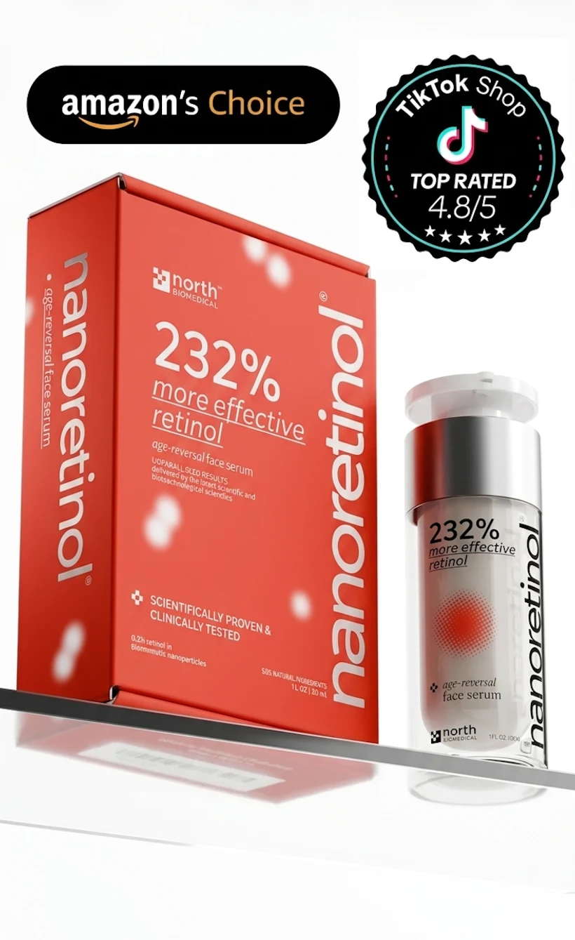
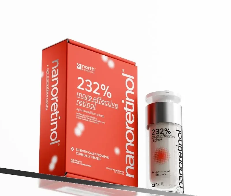
Nanoretinol® by North Biomedical® addresses this through lipid nanoparticle encapsulation that delivers 0.2% retinol via biomimetic carriers. The controlled-release mechanism means retinol reaches melanocyte-adjacent cells gradually rather than in a single irritating burst. Clinical data demonstrates +232% more effective collagen recovery and +73% more effective elastin recovery compared to conventional retinol, with significantly less irritation [6].
For the hands specifically, the water-based gel formulation absorbs quickly without leaving a greasy residue — a practical consideration for a product you will apply to hands that need to function immediately after application. The 99% natural ingredient profile avoids the synthetic irritants that make conventional retinol formulations particularly harsh on repeatedly washed hand skin.
A Realistic Protocol for Fading Age Spots on Hands
- Morning: Apply vitamin C serum (10–20% L-ascorbic acid) to the backs of hands, followed by a broad-spectrum SPF 50 sunscreen. Reapply sunscreen after every hand wash.
- Evening: Apply retinoid treatment to the backs of hands. Start every other night and increase frequency as tolerated.
- Weekly: Consider adding a gentle glycolic acid treatment (8–10%) once weekly to accelerate surface cell turnover.
- Ongoing: Wear UV-protective driving gloves for daily commutes. This single habit eliminates one of the largest sources of cumulative hand UV exposure.
Visible improvement typically begins after 8–12 weeks of consistent retinoid use, with progressive fading continuing over 6–12 months. Complete elimination of established solar lentigines through topicals alone may not be achievable — professional treatments may be needed for deep or long-standing lesions — but meaningful lightening and prevention of new spots is a realistic and evidence-supported outcome.
Age spots on hands are a permanent record of cumulative sun exposure — but that record can be partially rewritten with consistent treatment and, more importantly, the UV protection habits that prevent new chapters from being added.
References
-
Mardani N, Ghassemi M, Reza Safaei Naraghi Z, et al. Treatment of Solar Lentigines: A Systematic Review of Clinical Trials. J Cosmet Dermatol. 2025;24(3):e70133. doi:10.1111/jocd.70133
-
Fleischer AB Jr, Schwartzel EH, Colby SI, Altman DJ. The combination of 2% 4-hydroxyanisole (mequinol) and 0.01% tretinoin is effective in improving the appearance of solar lentigines. J Am Acad Dermatol. 2000;42(3):459-467. doi:10.1016/S0190-9622(00)90219-6
-
Draelos ZD. Novel approach to the treatment of hyperpigmented photodamaged skin: 4% hydroquinone/0.3% retinol versus tretinoin 0.05% emollient cream. Dermatol Surg. 2005;31(7 Pt 2):799-804. doi:10.1111/j.1524-4725.2005.31723
-
Varani J, Dame MK, Rittie L, et al. Vitamin A antagonizes decreased cell growth and elevated collagen-degrading matrix metalloproteinases and stimulates collagen accumulation in naturally aged human skin. J Invest Dermatol. 2000;114(3):480-486. doi:10.1046/j.1523-1747.2000.00876.x
-
Ortonne JP, Pandya AG, Lui H, Hexsel D. Treatment of solar lentigines. J Am Acad Dermatol. 2006;54(suppl 2):S262-S271. doi:10.1016/j.jaad.2005.12.043
-
North Biomedical LLC. “Nanoretinol® vs. Conventional Retinol: Efficacy in Collagen and Elastin Recovery.” Clinical Study Summary, 2024.