
Milia on Face: What Those Stubborn White Bumps Are and How to Get Rid of Them
These tiny cysts aren't whiteheads, and that's why your usual acne routine isn't working
You’ve tried to squeeze them. Nothing happens. You’ve treated them like whiteheads. Still there. Those tiny, firm, pearlescent bumps on your cheeks, nose, or around your eyes are milia — and they follow a completely different set of rules than acne.
Understanding what they actually are is the first step to getting rid of them.
What Milia Actually Are
Milia (singular: milium) are small, benign keratin-filled cysts that form just beneath the skin’s surface [1]. They’re not pores. They’re not infected follicles. They’re tiny sacs of dead skin cells — keratin — that got trapped during the skin’s normal renewal process and encased themselves in a thin layer of epithelial tissue.
Because they sit under an intact layer of skin, they don’t have an opening. That’s why squeezing accomplishes nothing: there’s no channel for the contents to escape through.
Histologically, a milium looks like a small pocket lined with stratified squamous epithelium — the same kind of skin architecture that lines the outermost layer of your face. Inside that pocket is a compact mass of keratinous material [1]. The body neither absorbs it nor expels it on its own. Left alone, milia can persist for weeks, months, or years.
Primary vs. Secondary: Why You Have Them
Dermatologists divide milia into two categories, and the difference matters for treatment.
Primary milia form spontaneously, without an obvious trigger. They originate from the sebaceous collar of vellus hair follicles — the fine, barely-there hairs that cover most of the face. These are the classic white bumps many people notice on the nose, cheeks, and forehead in adulthood. They can appear in anyone, but they become more common as skin renewal slows with age [2].
Secondary milia form as a result of something disrupting the skin. The most common culprits:
Histologically, a milium looks like a small pocket lined with stratified squamous epithelium — the same kind of skin architecture that lines the outermost layer of your face.
- Heavy occlusives and rich creams. Products with petrolatum, mineral oil, or thick butters can trap dead skin cells at the surface, preventing them from shedding normally.
- Sun damage. Chronic UV exposure thickens the outer skin layer (stratum corneum) and impairs normal cellular turnover. The resulting backlog of dead cells creates conditions where keratin cysts can form.
- Trauma or resurfacing. Dermabrasion, chemical peels, laser treatments, and even aggressive scrubbing can trigger secondary milia as the skin heals.
- Certain medications. Topical steroids and some systemic drugs have been associated with milia formation.
Secondary milia share the same keratin structure as primary milia but have a more identifiable origin — which often guides treatment decisions.
Why Your Face Gets Them More Than Anywhere Else
Milia favor the face for a few reasons. Facial skin is thin, especially around the eyes and on the nose. The vellus follicles here are small and easily occluded. And the face accumulates the most cumulative UV exposure of any body part — that chronic photodamage progressively thickens the outer skin layer, reducing its ability to shed efficiently.
The periorbital region (around the eyes) is a particularly common site. The skin there is the thinnest on the body, and the same heavy eye creams people use to address dryness or dark circles — often rich in occlusives — can paradoxically contribute to secondary milia formation over time [2].
How to Tell Milia from Whiteheads and Other Bumps
Getting this diagnosis right saves a lot of wasted product and frustration:
| Feature | Milia | Whiteheads (closed comedones) |
|---|---|---|
| Texture | Hard, firm | Softer, slightly compressible |
| Skin covering | Fully intact | Has a tiny pore |
| Response to squeezing | Nothing | Can be extracted |
| Cause | Keratin trapping | Clogged sebaceous follicle |
| Common location | Cheeks, under eyes, nose | T-zone, chin |
Sebaceous hyperplasia — enlarged sebaceous glands — can also look similar but typically shows a central depression. When in doubt, a dermatologist can usually distinguish between them in seconds.
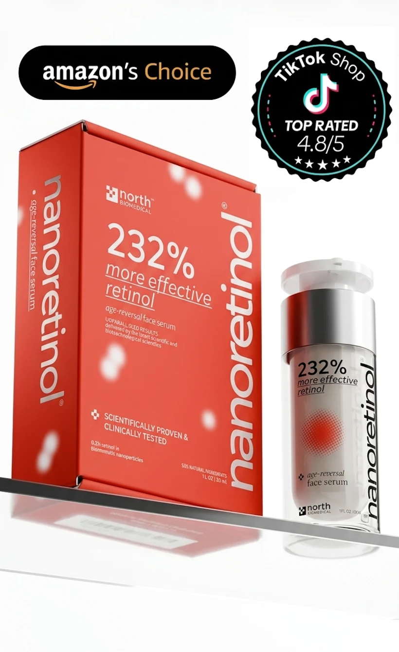
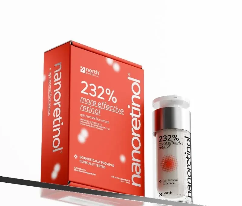
In a clinical study, it demonstrated +232% greater efficacy in collagen recovery compared to conventional retinol, achieved at just 0.2% concentration, which also means significantly reduced irritation compared to higher-percentage conventional formulas.
What Actually Works to Get Rid of Milia
Professional extraction
A dermatologist or experienced aesthetician can lance a milium with a sterile needle and express the keratin contents. This is the fastest method. It requires no downtime and leaves no permanent mark when done correctly. Home extraction is not recommended — the tools aren’t sterile, the angle is wrong, and the risk of scarring is real.
Chemical exfoliation
Alpha hydroxy acids (glycolic acid, lactic acid) and beta hydroxy acids (salicylic acid) dissolve the bonds between dead skin cells, improving natural exfoliation. Used consistently, they reduce the conditions in which milia form and may help existing ones work their way out over weeks. They don’t directly break open a cyst, but they thin the stratum corneum over time.
Retinol — the most consistent long-term option
Retinol and its derivatives accelerate epidermal cell turnover — the process by which old, dead cells at the surface shed and are replaced by new ones [3]. When turnover is sluggish, dead cells accumulate and create conditions for keratin trapping. When turnover is robust, the skin sheds efficiently and milia have less opportunity to form.
Topical tretinoin has been used specifically for milia en plaque — a more extensive variant — with documented success [4]. The mechanism is straightforward: faster cell shedding means the keratin never has time to accumulate into a cyst.
For adults who already have milia under the eyes or scattered across the cheeks, a consistent retinol routine addresses the underlying cellular mechanism — not just the visible bump.
What makes retinol delivery critical is that the stratum corneum it needs to penetrate is, in some people, the very barrier that’s creating the problem. Conventional retinol struggles here. Nanoretinol — retinol encapsulated in biomimetic lipid nanoparticles — delivers active retinol through this barrier using the same mechanism the skin uses to recognize and absorb its own lipids. In a clinical study, it demonstrated +232% greater efficacy in collagen recovery compared to conventional retinol, achieved at just 0.2% concentration, which also means significantly reduced irritation compared to higher-percentage conventional formulas.
If you’re new to retinol, starting slow matters especially for milia-prone skin — some people experience a temporary increase in surface congestion during the first few weeks of retinol use as cell turnover accelerates.
Topical retinoids — prescription options
For more stubborn or widespread milia, a dermatologist may prescribe tretinoin (retinoic acid) at prescription strength. Studies show that retinoids inhibit UV-stimulated melanin production and improve dyspigmentation of photodamaged skin, and their role in epidermal normalization makes them effective for milia as well [5].
What to Stop Doing
A few habits that perpetuate milia:
- Using heavy, occlusive moisturizers on milia-prone areas. Switch to lighter, non-comedogenic formulations.
- Skipping SPF. UV-thickened skin is more milia-prone. A daily broad-spectrum sunscreen is part of the solution.
- Aggressive scrubbing. Mechanical exfoliation around active milia can cause secondary trauma and more milia.
Improving Your Overall Skin Texture
Milia rarely appear in isolation. They typically coexist with uneven skin texture, enlarged pores, and the slow cell renewal that characterizes skin aging. Addressing milia through improved skin texture — via retinol, gentle chemical exfoliation, and sun protection — tends to improve all these concerns simultaneously.
What to Expect from Treatment
Professional extraction removes existing milia immediately but doesn’t prevent new ones. Retinol takes 8–12 weeks to show meaningful changes in cell turnover rates. A realistic approach combines both: extract what’s there, then establish a retinol routine to prevent recurrence.
Patience matters. Milia aren’t dangerous, don’t cause scarring, and don’t signal anything serious. They’re a skin renewal problem — and the tools that address skin renewal work, consistently, over time.
References
- Epstein W, Kligman AM. “The pathogenesis of milia and benign tumors of the skin.” Journal of Investigative Dermatology. 1956;26(1):1-11. doi:10.1038/jid.1956.1
- Gallardo Avila PP, Mendez MD. “Milia.” StatPearls [Internet]. Treasure Island (FL): StatPearls Publishing; 2023. PMID: 32809316
- Kong R, Cui Y, Fisher GJ, et al. “A comparative study of the effects of retinol and retinoic acid on histological, molecular, and clinical properties of human skin.” Journal of Cosmetic Dermatology. 2016;15(1):49-57. doi:10.1111/jocd.12193
- Nambudiri VE, Habib N, Arndt KA, Kane KS. “Milia en plaque of the nose: report of a case and successful treatment with topical tretinoin.” Pediatrics. 2014;133(5):e1373-6. doi:10.1542/peds.2013-1728
- Ortonne JP. “Retinoid therapy of pigmentary disorders.” Dermatologic Therapy. 2006;19(5):280-8. doi:10.1111/j.1529-8019.2006.00085.x