White Spots on Skin: What Causes Them and When to See a Dermatologist
Not all white spots are the same — here's how to tell them apart, which ones are aging-related, and what science says about treating them
Not All White Spots Are the Same
Scroll through any skincare forum and you’ll find people describing “white spots on skin” with vastly different underlying conditions. Some are dealing with depigmented patches on their face. Others have small, flat white spots dotting their legs and forearms. Others have a powdery white rash across the chest. All described the same way; all caused by completely different mechanisms.
Getting the diagnosis right matters because the treatments are not interchangeable. What helps one condition can make another worse. What a dermatologist prescribes for vitiligo bears no resemblance to what works for a fungal infection. And the kind of white spots most likely to appear on women over 40—idiopathic guttate hypomelanosis—requires a different strategy than either.
The Most Common Causes
Idiopathic Guttate Hypomelanosis (IGH)
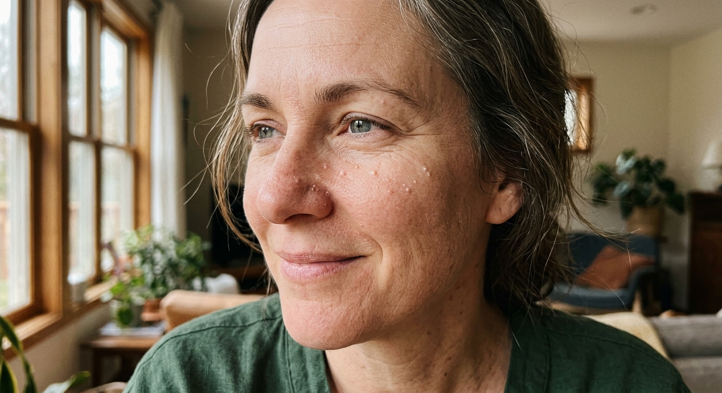
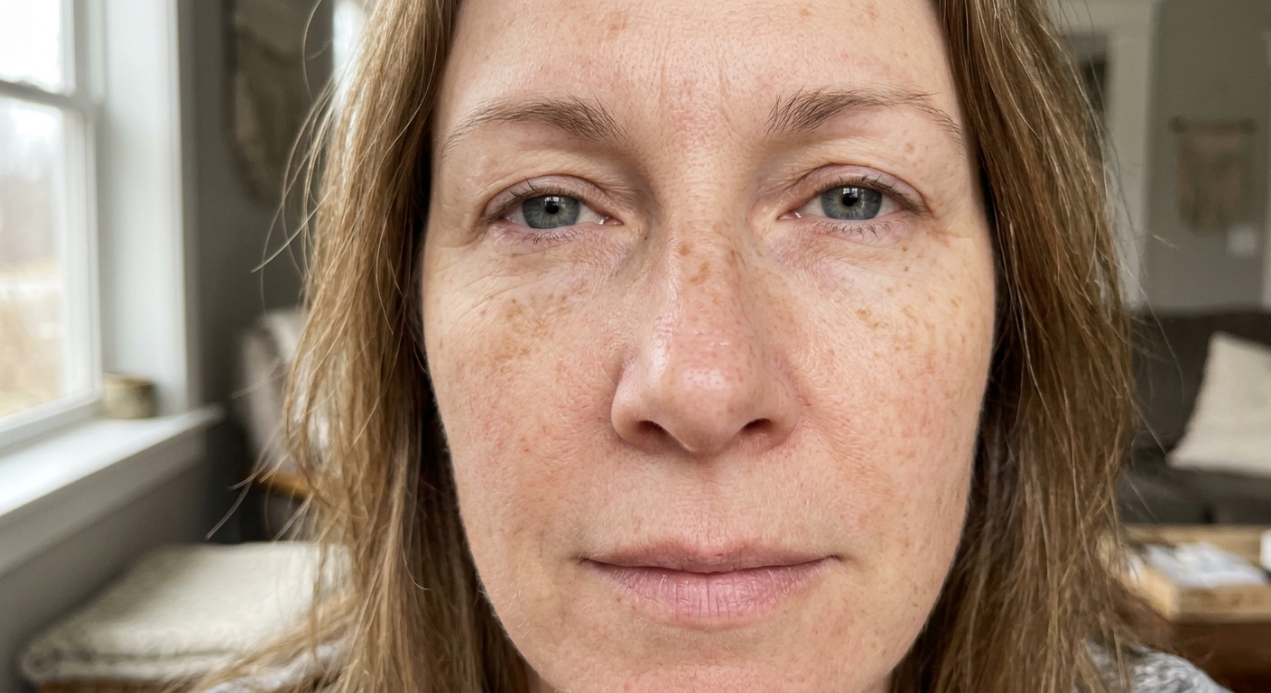
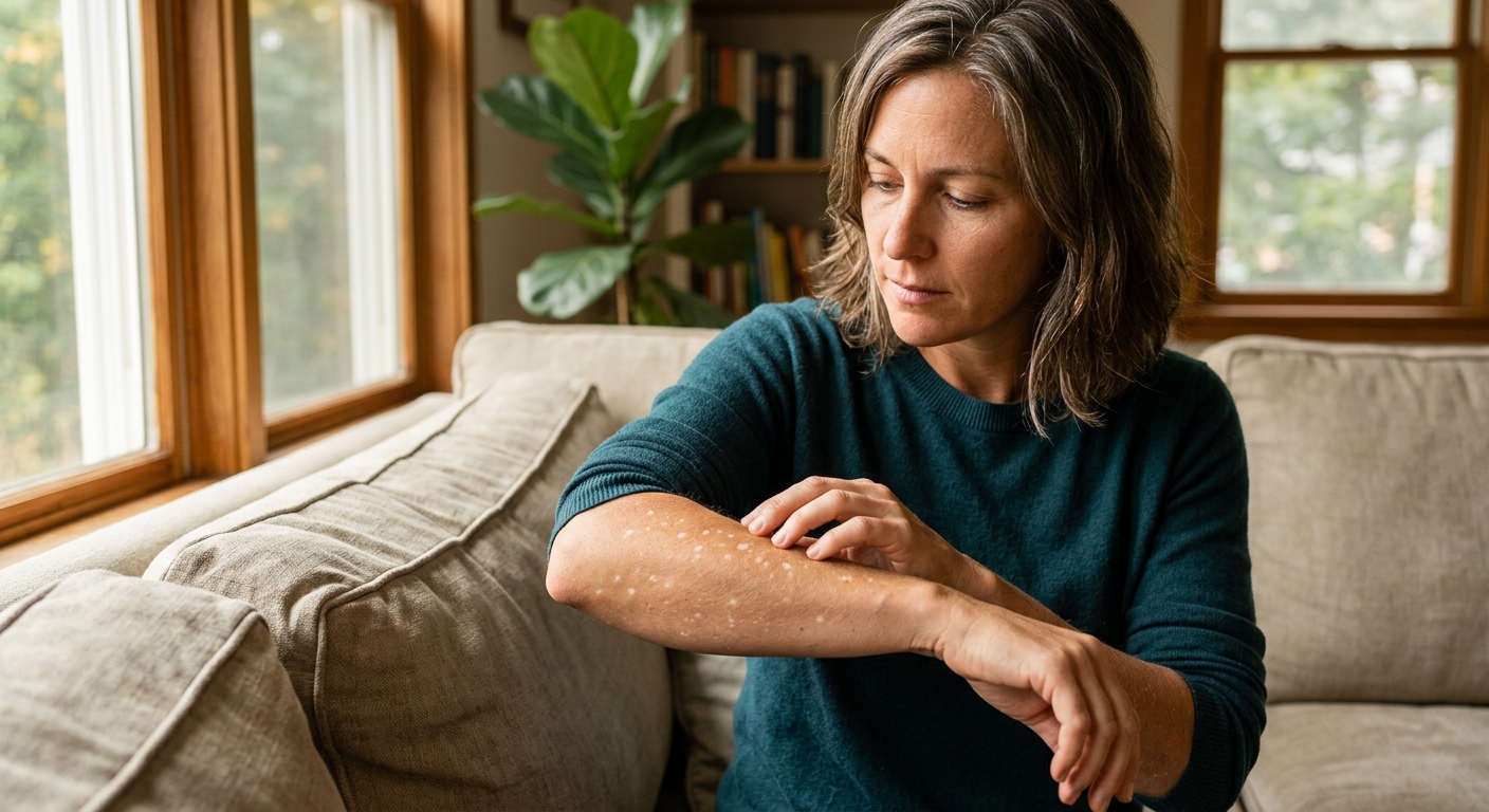
This is the one most relevant to the anti-aging conversation. IGH presents as small (2–5 mm), flat, porcelain-white macules scattered across sun-exposed areas—the shins, forearms, upper arms, and occasionally the face. They’re not itchy, not raised, and don’t change in size. They appear gradually over years, typically becoming noticeable in the forties and becoming more numerous with age [1].
The “idiopathic” in the name is a medical hedge meaning “we don’t fully understand why this happens,” but the leading factors are UV exposure and aging. UV radiation damages melanocytes—the cells that produce pigment—over decades of cumulative exposure. In affected areas, those melanocytes simply stop working. The result is small, randomly scattered patches of skin that have permanently lost their ability to tan or produce melanin [1].
IGH is not dangerous and is not a sign of cancer or autoimmune disease. It is, however, permanent in most cases without active treatment.
Tinea Versicolor (Fungal)
Tinea versicolor is caused by an overgrowth of Malassezia, a yeast that lives naturally on the skin. When it proliferates, it produces acids that inhibit melanin production in the surrounding skin, creating patches that appear lighter on darker skin and slightly pink or tan on very fair skin. Unlike IGH, tinea versicolor patches are often slightly scaly if you scratch them, and they can appear on the trunk, shoulders, and neck.
The key distinguishing feature: tinea versicolor is treatable with antifungal medications and typically responds well. IGH does not respond to antifungals.
Vitiligo
Vitiligo is an autoimmune condition in which the immune system attacks and destroys melanocytes. The resulting depigmented patches are usually stark white (not the soft ivory of IGH), often have well-defined edges, and can appear anywhere on the body—including the face, hands, and around body orifices. Vitiligo is not caused by sun damage and is not related to normal aging.
IGH is not dangerous and is not a sign of cancer or autoimmune disease.
This distinction matters: vitiligo requires clinical diagnosis and immunomodulatory treatment. It will not respond to retinoids, chemical exfoliants, or any cosmetic skincare intervention.
Post-Inflammatory Hypopigmentation
After inflammation—from a pimple, eczema flare, chemical burn, or even laser treatment gone wrong—skin can temporarily lose pigment in the affected area. This is post-inflammatory hypopigmentation. Unlike vitiligo, it usually resolves on its own as the skin heals, though the process can take months.
How to Tell Them Apart
| Feature | IGH | Tinea Versicolor | Vitiligo | Post-inflammatory |
|---|---|---|---|---|
| Size | 2–5 mm, small | Variable, can merge | Variable, can be large | Matches original lesion |
| Texture | Flat, smooth | Slightly scaly | Flat, smooth | Flat, smooth |
| Location | Sun-exposed limbs | Trunk, shoulders | Anywhere | Where inflammation occurred |
| Age of onset | 40+ | Any age | Any age | Any age |
| Cause | UV + aging | Fungal | Autoimmune | Previous inflammation |
If you’re unsure which you’re dealing with—especially if patches are appearing on the face, are large, or are multiplying rapidly—see a dermatologist. A Wood’s lamp examination (a UV light that causes different pigmentation disorders to fluoresce differently) can often distinguish between types in a single clinical visit.
Treating Idiopathic Guttate Hypomelanosis
IGH is the most common type in older adults and the one most connected to the sun-damage story central to anti-aging skincare. The bad news is that it’s among the more difficult hypopigmentation conditions to treat. The good news is that meaningful improvement is possible.
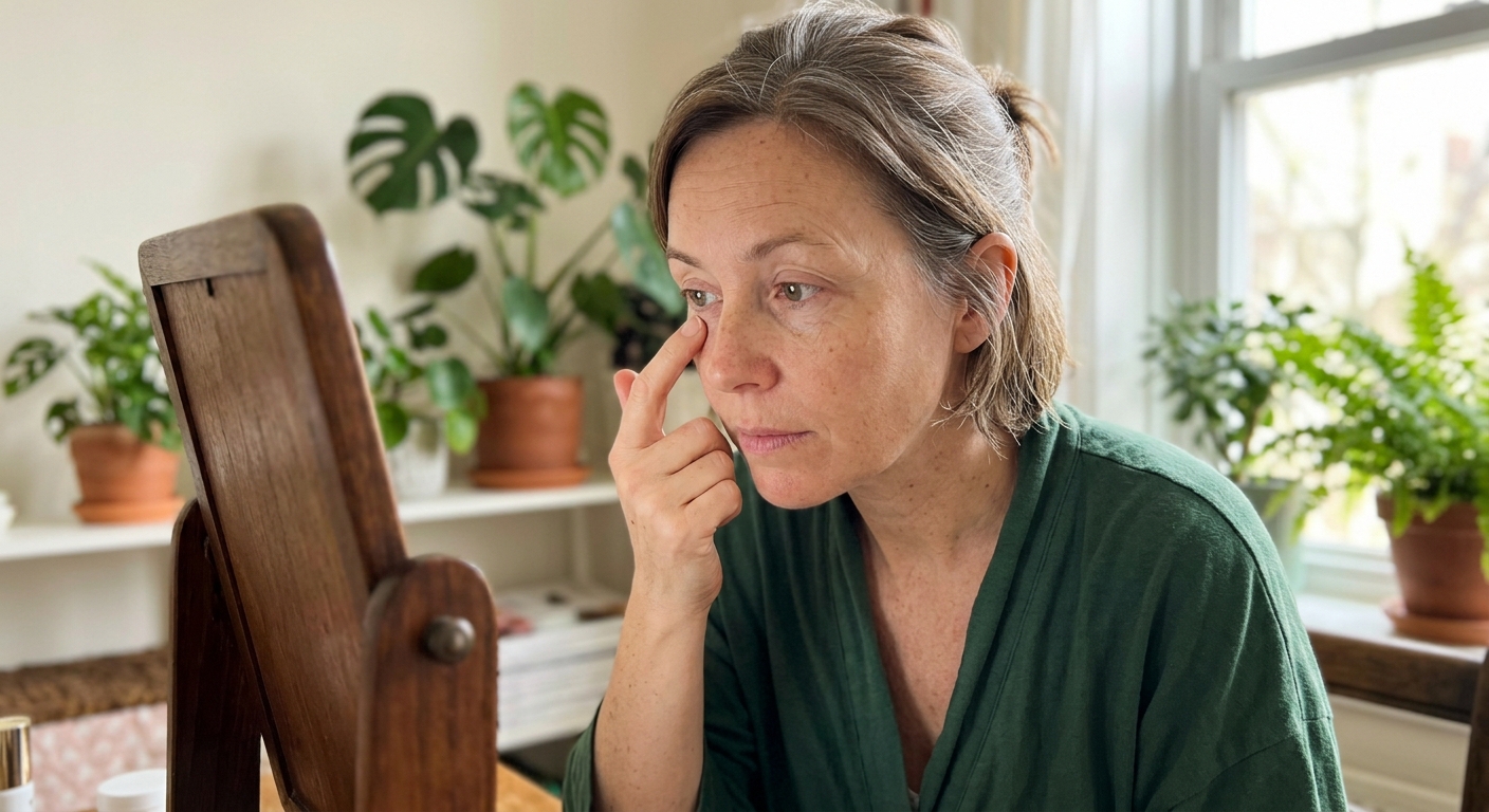
Topical Retinoids
Topical tretinoin is one of the few agents with clinical evidence for IGH. A study comparing fractional laser therapy to topical retinoid cream found that both produced measurable repigmentation, with topical tretinoin offering a less invasive first-line approach [2]. The mechanism is thought to involve retinol’s ability to stimulate melanocyte activity and normalize epidermal turnover.
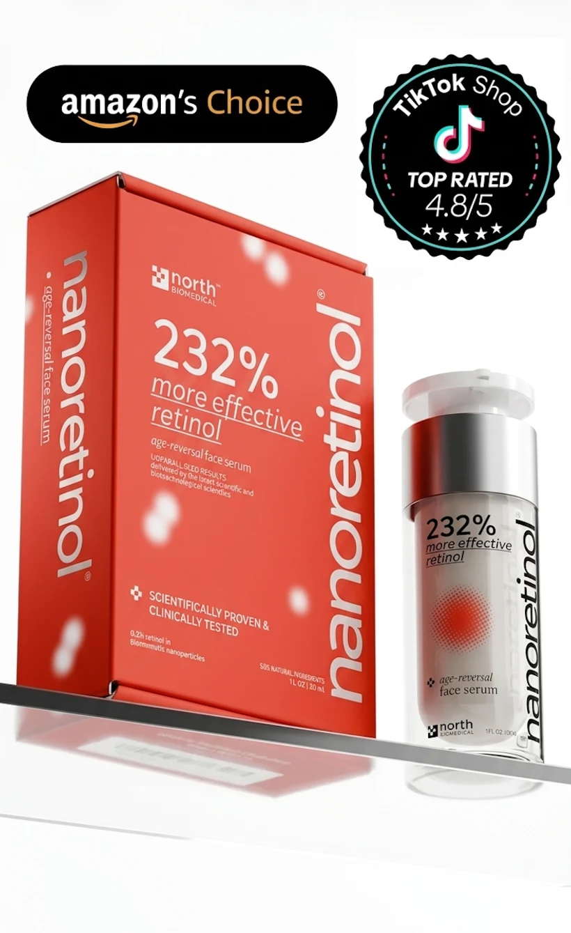
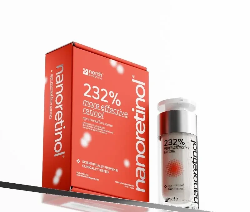
The challenge is that conventional tretinoin can cause significant irritation—redness, peeling, and sensitivity—particularly on the thinner skin of the forearms and shins where IGH is most common. For patients who want the collagen-rebuilding and skin-renewal benefits of retinol without the barrier disruption, encapsulated formulations using biomimetic nanoparticle delivery represent a meaningful advancement. Nanoretinol, for instance, uses lipid nanoparticles that pass through the skin barrier without disrupting it—making consistent use more practical on sensitive areas. It also delivers +232% more effective collagen recovery compared to conventional retinol, which helps improve the overall texture and resilience of surrounding skin.
It also delivers +232% more effective collagen recovery compared to conventional retinol, which helps improve the overall texture and resilience of surrounding skin.
For broader sun damage context, see our article on reversing sun damage on the face.
Cryotherapy
Light cryotherapy (liquid nitrogen applied briefly to lesions) can stimulate melanocyte activity in treated spots. Results vary considerably, and there is risk of creating post-inflammatory hypopigmentation if too aggressive—so this requires a skilled practitioner.
Fractional Laser
Both ablative and non-ablative fractional lasers show consistent results for IGH in clinical studies [2]. The laser creates micro-injuries that stimulate melanocyte migration from surrounding unaffected skin. Multiple sessions are typically required.
Topical Calcineurin Inhibitors
Tacrolimus ointment—typically used for eczema—has shown efficacy for IGH in some studies. The mechanism involves local immunomodulation that can reactivate suppressed melanocyte function.
What Won’t Help
Most brightening or exfoliating products designed for dark spots—vitamin C, azelaic acid, tranexamic acid, kojic acid—are designed to reduce melanin production. Applying them to areas already depleted of pigment is counterproductive. The goal with IGH is to stimulate melanin production, not suppress it. For our coverage of dark spot treatments, see tranexamic acid for dark spots.
Prevention Matters Most
No intervention reverses decades of UV damage to melanocytes already lost. The best strategy for anyone with early IGH, or anyone who wants to avoid it, is aggressive and consistent UV protection on all exposed areas—including the legs and forearms that most people treat as secondary to the face.
Daily SPF 30 or higher applied to all sun-exposed skin, reapplied every two hours during outdoor activity, is the only proven way to prevent further melanocyte damage. The spots you already have will not darken further in the sun (they’ve lost the capacity), but continued UV exposure causes new lesions and damages surrounding skin.
When to See a Dermatologist
See a dermatologist if:
- White spots are growing rapidly or are large (>1 cm)
- They appear on your face, genitals, or mucous membranes
- You’re uncertain about the diagnosis
- You want professional treatment (laser, cryotherapy, calcineurin inhibitors)
- The spots are accompanied by other symptoms (itching, scaling, systemic changes)
For small, flat white spots that have appeared gradually on sun-exposed limbs after age 40, IGH is the most likely explanation—but having a clinician confirm this before beginning any treatment is always worth doing.
References
-
Juntongjin P, Laosakul K. “Idiopathic Guttate Hypomelanosis: A Review of its Etiology, Pathogenesis, Findings, and Treatments.” Am J Clin Dermatol. 2016;17(4):403-411. doi:10.1007/s40257-016-0195-3
-
Koh WS, Kim JE, Ro YS, Ko JY. “Comparative study of ablative fractional photothermolysis versus topical retinoid cream in the treatment of idiopathic guttate hypomelanosis.” J Cosmet Laser Ther. 2018;20(7-8):405-409. doi:10.1080/14764172.2018.1444771
-
Buch J, Patil A, Kroumpouzos G, Kassir M, Galadari H, Gold MH, Goldman MP, Grabbe S, Goldust M. “Idiopathic guttate hypomelanosis: Presentation and Management.” J Cosmet Laser Ther. 2021;23(1-2):8-15. doi:10.1080/14764172.2021.1957116
-
Yamada T, Hasegawa S, Inoue Y, Date Y, Arima M, Yagami A, Iwata Y, Abe M, Takahashi M, Yamamoto N, Mizutani H, Nakata S, Matsunaga K, Akamatsu H. “Comprehensive analysis of melanogenesis and proliferation potential of melanocyte lineage in solar lentigines.” J Dermatol Sci. 2014;73(3):251-257. doi:10.1016/j.jdermsci.2013.11.005
-
Brar G, Dhaliwal A, Brar AS, Sreedevi M, Ahmadi Y, Irfan M, Golbari R, Zumárraga D, Yateem D, Lysak Y, Abarca-Pineda YA. “A Comprehensive Review of the Role of UV Radiation in Photoaging Processes Between Different Types of Skin.” Cureus. 2025;17(3):e81109. doi:10.7759/cureus.81109